

 Despina Petrushevska

Biopsychosocial questionnaire
What is a biopsychosocial questionnaire? A biopsychosocial questionnaire is a st...

1
Last Updated: Nov 18, 2026
Free PDF 2 pages, print-ready Clinician-reviewed
A blood flow chart through the body shows how blood moves from the heart to the lungs, around the body and back again. Deoxygenated blood enters the right atrium, passes to the right ventricle and travels through the pulmonary artery to the lungs. Oxygenated blood returns to the left atrium, fills the left ventricle and leaves through the aorta to reach every tissue. This free two-page chart labels each chamber, valve and major vessel in order. Nurse educators, students and practice teams can use it as one shared reference for explaining the route.
A two-page chart with a labeled diagram of both circuits, a ten-step pathway table and a right versus left ventricle comparison. It ends with six check-your-understanding questions and space for notes.
Start from scratch, or upload this PDF or any form you already use, and Pabau's AI Form rebuilds it with tick boxes, text fields and a signature. Attach it to the treatment once, and every patient gets it on time.
AI Form rebuilds the consent, history and aftercare forms you already use. You review each one, then publish.
Patients complete forms on their phone before they arrive, so the front desk stops retyping paper.
Every signed form is versioned and attached to the treatment record, ready when you need it.
Set how long a consent stays valid. Returning patients skip forms they have already signed.
Supports HIPAA and GDPR compliance AES-256 encryption 4.8 on G2 4.7 on Capterra Trusted by 4,000+ practices worldwide

Blood travels through two linked loops, the pulmonary circuit to the lungs and the systemic circuit to the rest of the body.
Deoxygenated blood enters the right atrium through the superior and inferior venae cavae.
Oxygenated blood leaves the left ventricle through the aorta to reach every tissue.
The pulmonary artery and pulmonary veins reverse the usual oxygen pattern of arteries and veins.
Use the chart for patient education, staff induction and anatomy teaching.
The circulatory system is a closed network of heart, blood vessels and blood that delivers oxygen and nutrients to tissues and removes waste. The NHLBI’s guide to heart blood flow traces the same route the chart follows. Oxygen-poor blood enters the right atrium, and the right ventricle pumps it to the lungs. Oxygen-rich blood then returns to the left heart before the aorta carries it to the body.
That route runs as two linked circuits driven by the same heartbeat. The pulmonary circuit carries blood between the heart and lungs, while the systemic circuit carries it between the heart and the rest of the body.
Blood flows through the heart in a fixed sequence, and the chart follows that order. Each stage is one step in a single loop, so the arrows never change direction. For a closer look at the chambers themselves, pair it with a labeled heart diagram.
Pulmonary circulation is the short loop that moves blood to the lungs for gas exchange. It starts in the right ventricle and ends at the left atrium. The chart colors the outbound half blue for deoxygenated blood and the returning pulmonary veins red for oxygenated blood.
Systemic circulation is the longer loop that delivers oxygenated blood to every tissue. It starts in the left ventricle and returns to the right atrium. Capillaries at the tissue level exchange oxygen and nutrients before venous blood returns to the heart.
The chart labels the main vessels and structures you need to read the diagram accurately. Use the table to check each label against its role.
Two rows in that table catch learners out. The pulmonary artery is the only artery that carries deoxygenated blood, and the pulmonary veins are the only veins that carry oxygenated blood. Artery and vein describe direction relative to the heart, so oxygen content plays no part in the name.
Heart valves keep blood moving in one direction through each chamber. The tricuspid valve sits between the right atrium and right ventricle, and the mitral valve sits between the left atrium and left ventricle. The pulmonary and aortic valves control exit from the two ventricles.
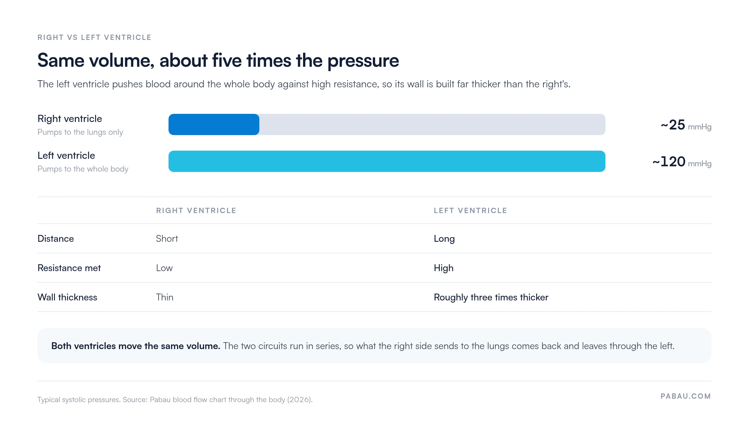
The two ventricles pump the same volume of blood with each beat, because the circuits run in series. What the right ventricle sends to the lungs must come back and leave through the left. The difference between them is pressure.
The right ventricle only has to reach the lungs, a short, low-resistance trip. The left ventricle drives blood around the whole body against far higher resistance. The chart puts typical systolic pressure at about 25 mmHg on the right and about 120 mmHg on the left. That workload is why the left wall is roughly three times thicker.
That volume-versus-pressure distinction is the bridge to cardiac output. Learners who want to put numbers on each beat can work through the stroke volume equation next.
Some circulation routes sit outside the basic two-circuit model, and the chart leaves them out. Cover them separately when a learner asks, so nobody confuses them with the main systemic loop.
Coronary blood supply is separate from the pulmonary circuit, which serves gas exchange only. Learners often mix these up, so describe the coronary arteries as a supply to the heart wall, not a route to the lungs.
The chart works best as a shared reference rather than a standalone handout. Use it at the point where the question comes up, so the explanation matches what the patient or learner is looking at.
The chart explains anatomy and flow only. It gives no diagnostic or treatment guidance, so any question about a specific patient belongs with the treating clinician. When the conversation moves from anatomy to symptoms, a structured circulation assessment is the clinical next step.
The two-page PDF traces the full pathway from the body tissues through the heart and lungs and back again. It is free to download and print for teaching and patient information use.
Most practices hand out a printed chart during the appointment, then explain circulation from scratch while the patient reads. The explanation competes with the clock, and nobody records which patient received which handout.
Pabau, the practice management platform we build, sends the chart with pre-appointment forms so the patient reads it at home. The digital form lands on the patient’s record once it is completed, which shows the team who has seen the material. For practices that run that workflow across a whole patient list, our primary care practice software keeps forms, bookings and notes in one system.
The appointment then starts from shared ground. The clinician spends the time on the patient’s own questions rather than on the anatomy basics.

Pabau’s digital forms deliver reference material like this chart ahead of the appointment and store the completed form on the patient’s record.

Teach circulation as one loop with two pumps, and most of the confusion learners bring to it disappears. Start at the body tissues, follow the arrows clockwise, and stop at the two exceptions where arteries and veins swap their usual oxygen content.
The chart covers anatomy and flow. Symptoms and diagnosis still need a clinician, so keep the handout as a starting point for that conversation. Book a demo to see how Pabau sends education material like this to patients before they arrive.

Want the chambers in more detail? Heart diagram labels each chamber, valve and vessel on a single printable page.
Testing what learners remember? ECG quiz checks how well staff read the heart’s electrical activity.
Teaching cardiac output next? Cardiac output formula shows how heart rate and stroke volume combine.
Explaining blood groups as well? Blood type chart sets out donor and recipient compatibility.
Referring a patient for imaging? Echocardiogram test explains what the scan shows about heart structure and function.
A blood flow chart through the body is a diagram that traces how blood moves through the heart, lungs and systemic circulation. It labels the chambers, valves and major vessels in sequence.
Common signs include cold extremities, numbness, slow-healing wounds and pain on walking. These are symptoms to raise with a clinician, and the chart does not diagnose them.
Pulmonary circulation carries blood between the heart and lungs for gas exchange. Systemic circulation carries oxygenated blood from the heart to the body and returns deoxygenated blood to the right atrium.
The heart valves open and close to keep blood moving forward through each chamber. They prevent blood from flowing back into the chambers it has just left.