



GLP-1 diet plan
A GLP-1 diet plan is a structured eating framework for patients taking semagluti...

2
Last Updated: Oct 13, 2026
The valsalva test asks a patient to bear down against a closed airway, and the pressure that builds does the diagnostic work. That strain raises pressure inside the chest, the abdomen and the spinal canal at once.
One fact governs all three. A positive result carries weight, but a negative one does not exclude the pathology. Miss that and you order the wrong next step.
Here is how to perform the maneuver, read its four phases, and record the finding so a colleague can use it.

The valsalva test does three separate jobs: spinal provocative testing, SVT termination, and autonomic assessment.
Specificity sits near 94% and sensitivity near 22%, so a positive result confirms while a negative one leaves the question open.
A Valsalva ratio of 1.5 or above is normal in adults, and below 1.2 is clearly abnormal.
The modified maneuver converted 43% of SVT episodes in the REVERT trial, against 17% for standard technique.
Glaucoma, retinal detachment, recent myocardial infarction, and severe aortic stenosis are absolute contraindications.
The valsalva test is a forced expiration against a closed airway. Most clinicians get there by asking the patient to pinch the nose, shut the mouth and blow hard. Bearing down as if straining at stool works just as well.
The rise in intrathoracic and intra-abdominal pressure is the mechanism. It compresses venous structures in the chest and the spinal canal, shifts cardiac preload and afterload, and triggers the baroreflex. Each clinical use then draws on a different part of that cascade.
For orthopedic and physical therapy exams, raised intrathecal pressure is the variable that matters. It aggravates a nerve root that is already compressed, which is what brings radicular symptoms back.
In cardiology, the response pattern during and after the strain either exposes autonomic damage or breaks a re-entrant tachycardia outright. So the first decision is which question you are asking, rather than how to run the maneuver.
Each use draws on a different part of the pressure change, so the protocols diverge quickly.
Which indication applies decides position, duration and interpretation. A 15-second seated strain for a lumbar disc patient has little in common with a 40 mmHg strain held against a manometer. The grid below sets the three protocols side by side.
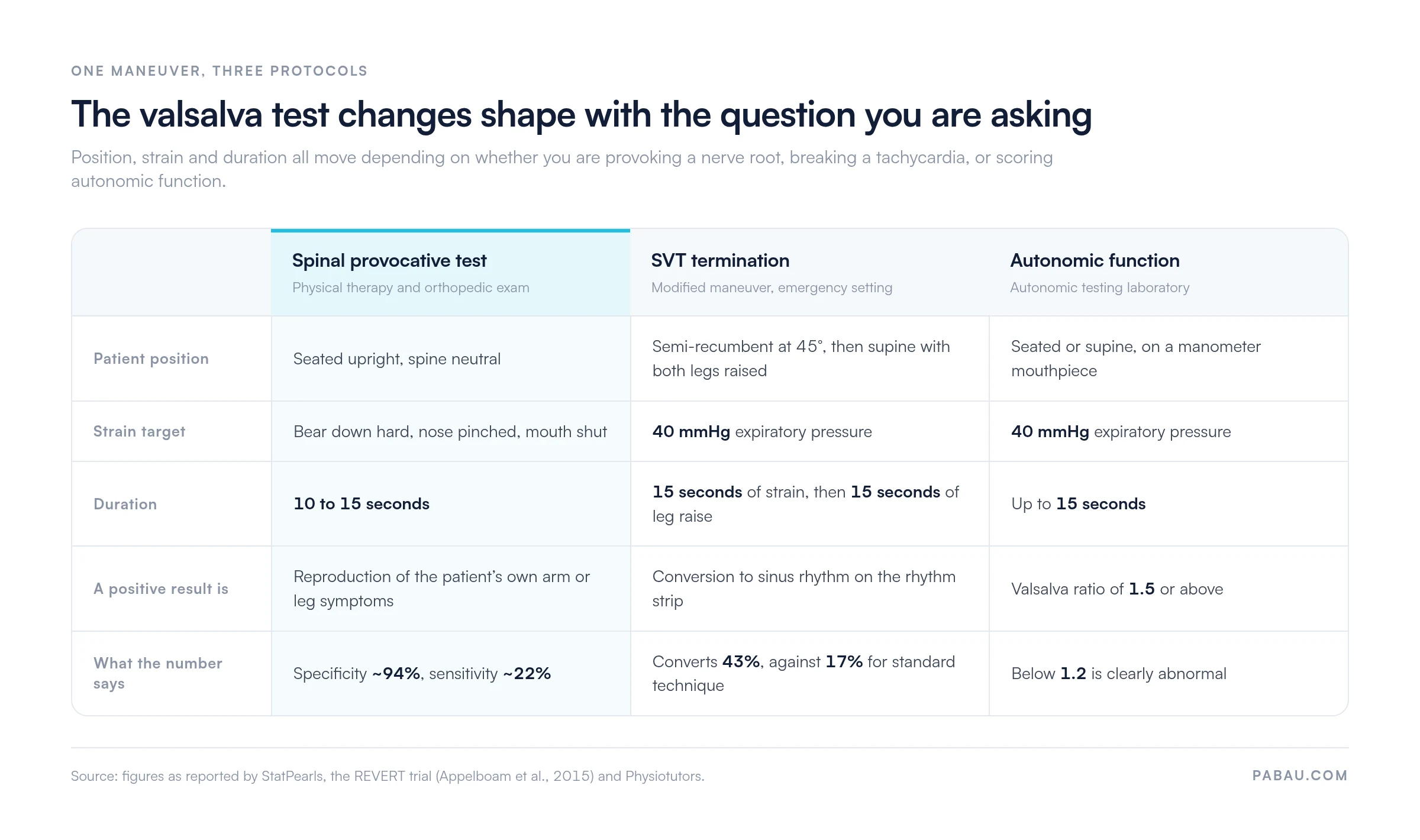
The technique below covers spinal provocative testing and general clinical use. The SVT and autonomic variants differ, and both are covered further down.
Ten to 15 seconds is the usual duration for provocative spinal testing. In a formal autonomic laboratory the patient holds 40 mmHg of expiratory pressure against a manometer mouthpiece. That runs for up to 15 seconds, with continuous ECG and blood pressure recording.
The modified valsalva maneuver adds a second stage to the standard strain, and the REVERT randomized controlled trial showed what that buys. Standard technique converted roughly 17% of episodes. The modified version converted 43%, according to the REVERT trial (Appelboam et al., The Lancet, 2015).
In emergency settings this is now the first protocol to try in stable SVT, ahead of drugs.
The cardiovascular response runs through four phases, and reading them is what turns the maneuver into an autonomic measurement. The table below sets out the hemodynamic change and the mechanism behind each one.
Phase IV carries most of the diagnostic weight. The overshoot and the reflex bradycardia behind it are the two events the Valsalva ratio is built from, as set out in StatPearls. When the overshoot fails to appear at all, sympathetic adrenergic failure is the usual explanation.
A positive valsalva test means three different things, and which one applies depends on why you ran it.
The ratio divides the longest R-R interval in the 30 seconds after the strain by the shortest R-R interval during it. Written out, that is the longest post-strain R-R over the shortest during-strain R-R.
In adults, 1.5 or above is generally normal, though reference ranges shift a little with laboratory protocol and age. Below 1.2 is widely treated as clearly abnormal and consistent with significant parasympathetic impairment.
Between 1.2 and 1.5 sits the borderline band. Read those results alongside the other autonomic indices, such as heart rate response to deep breathing and head-up tilt testing. A ratio that is effectively absent points to severe cardiovagal dysfunction, as seen in advanced diabetic autonomic neuropathy or multiple system atrophy.
The valsalva test is not a standalone diagnostic for cervical disc pathology, and its accuracy profile shows why.
Those figures come from Physiotutors’ clinical examination reference, and they describe a confirmatory tool rather than a screening one. A positive result in a patient you already suspect of cervical disc herniation moves the needle. A negative result in the same patient changes very little.
That low-sensitivity, high-specificity shape is why the maneuver belongs in an examination cluster. Pair it with Spurling’s test, upper-limb tension testing and a neurological exam, then let the pattern decide rather than any single item.
As a vagal maneuver for hemodynamically stable supraventricular tachycardia, the valsalva test is first-line. Raised pressure stimulates the baroreceptors, parasympathetic tone climbs, AV-node conduction slows, and the re-entrant circuit breaks.
Technique is what separates a one-in-six result from a four-in-ten one. The standard strain converts roughly one patient in six. The modified protocol described above converts around four in ten, on REVERT trial figures.
Before you try it, run through these decision points:
Document every Valsalva attempt for SVT with the time, the technique used, and the length of the strain. Add the rhythm before and after, plus how the patient responded. That record supports audit and gives the receiving team a clear handover if drugs become necessary.
Screen for these before any indication. Some are absolute, because the pressure spike can do irreversible harm. Others are relative, and they come down to a judgment call about risk against benefit.
Even a cleared patient carries a small risk, so know the pattern. Syncope is the commonest adverse event, driven by hypotension in phase II or an exaggerated phase IV bradycardia. Seat or lie the patient down, and stay with them throughout.
Valsalva retinopathy, meaning subconjunctival or retinal hemorrhage after a very forceful strain, is documented but uncommon. In patients with a history of transient ischemic attack or carotid disease, the hemodynamic swing carries a theoretical ischemic risk. That is rare in routine testing.
Stop immediately if the patient turns pale, becomes confused, or loses consciousness. Then manage the positional hypotension as you would after any faint.
Reference pages on the valsalva test tend to stop at interpretation. What decides whether the finding is worth anything six weeks later is the note you write about it.
A free-text line reading “Valsalva positive” cannot be audited, compared against last month’s exam, or handed to a colleague with any confidence. Named fields can. At minimum, the note records:
Serial comparison is where the structure pays off. A patient with progressive cervical disc disease may see three clinicians in six months, and only matched fields will show whether the picture is worsening. Practices shortlisting physiotherapy practice software usually test that first.
Audit is the second payoff. To learn how often a positive valsalva test led to an imaging referral, you need fields you can count, not paragraphs somebody has to read.
The valsalva test rarely decides anything on its own. Knowing what sits around it tells you when to add it, and when another test does more.
Most practices record a valsalva test the way they record the rest of the exam. It goes into a free-text box at the end of a busy clinic day. It reads fine at the time. Six weeks later, no one can say whether the strain lasted eight seconds or 15, or which arm the symptoms ran down.
Practice management software like Pabau replaces that box with named fields. Digital assessment forms let you build a provocative test template once, with position, technique, duration, result and symptom distribution captured separately. Every assessor then fills in the same form, so two exams six months apart can be compared directly.
Because those entries sit in the client record instead of a paragraph, they also count. You can pull how often a positive valsalva test led to imaging without reading a single note. And the next clinician to see that patient opens a shared physical therapy EMR, finding the same documented baseline rather than a colleague’s shorthand.


Pabau’s digital assessment forms and structured client records let physical therapy and sports medicine practices capture the same fields at every exam. Serial comparisons then hold up, and an audit takes minutes instead of an afternoon.

The valsalva test earns its place through specificity. Use it to firm up a suspicion you already hold, and read a negative result as absence of information rather than reassurance.
For SVT the question is settled. The modified protocol converts more than twice as many episodes as the standard strain, so make it the default in stable patients. For autonomic work, the Valsalva ratio gives you a number you can track over time.
The part that slips most easily is the record. A finding you cannot compare against the last exam has spent a clinician’s time and bought very little.
Structured records are what keep a provocative test useful at the next appointment. Book a demo to see how Pabau sets up assessment templates for physical therapy and sports medicine teams.

Working up a suspected cervical radiculopathy? Spurling’s test walks through the companion compression test you pair with this one, including how to score it.
Need to tell shoulder pain from a cervical referral? The Whipple test covers a shoulder special test that helps you place the source of the symptoms.
Assessing balance and joint position sense? Proprioception testing sets out the bedside methods and what each result tells you about neural control.
Looking for a wrist and thumb provocative test? The Finkelstein test explains the technique for De Quervain’s tenosynovitis and the errors that produce false positives.
They describe the same physical action. “Maneuver” usually refers to the act of straining, including its therapeutic use in SVT. “Test” refers to the diagnostic reading you take from it. Papers use both terms, so treat them as interchangeable when you search the literature.
Patients often use a gentle version to clear their ears, which is harmless for most people. A diagnostic strain is a different matter. Symptom reproduction and rhythm change need a clinician watching, and every contraindication still applies. Advise patients not to self-test for neck or back pain.
Yes, and that is a separate bedside use. Straining cuts venous return, which softens most systolic murmurs. The murmur of hypertrophic cardiomyopathy usually grows louder instead, because the outflow tract narrows as the ventricle empties. Cardiologists use that split to separate it from aortic stenosis.
Yes. Cardiovagal responses weaken as people get older, so a ratio that is acceptable at 70 would be low at 25. Autonomic laboratories therefore score results against age-matched reference values rather than one fixed cut-off. Treat a borderline figure in an older patient with that in mind.






