Chronic kidney disease nursing care plan
A chronic kidney disease nursing care plan is a structured document that guides ...

3
Last Updated: Oct 14, 2026

The valgus extension overload test is the primary clinical maneuver for posteromedial elbow impingement in overhead throwing athletes.
A positive result means posteromedial elbow pain reproduced while valgus stress is held and the elbow is passively extended from 70 degrees.
No published sensitivity or specificity value exists for the test, so the moving valgus stress test is used alongside it for UCL pathology.
Published recovery timelines conflict and are largely uncited, so plan around severity, imaging, and Pitch Smart workload limits.
Practice management software like Pabau helps sports medicine practices structure exam findings and track an athlete through each rehab phase.
The valgus extension overload test reproduces posteromedial elbow pain by applying valgus stress while you passively extend the elbow.
It is the maneuver practitioners reach for when valgus extension overload (VEO) syndrome is suspected in a thrower.
Posteromedial elbow pain in a baseball pitcher or cricket bowler is rarely straightforward, and several conditions share that anatomical territory. Getting the source wrong delays return to sport. The test is quick, needs no equipment, and narrows the differential when it is interpreted correctly.
This guide covers how to perform the test step by step and how to read the result. It also compares the test with the moving valgus stress test. Then it covers what comes next, from imaging and differential diagnosis to recovery timelines, rehab exercises, and the workload limits that reduce VEO risk.
The valgus extension overload test is an examination maneuver that reproduces posteromedial elbow pain through the last 70 degrees of elbow extension. It replicates the forces the elbow meets during the acceleration and deceleration phases of throwing. The aim is to provoke olecranon impingement against the posteromedial olecranon fossa.
That is the same mechanism driving osteophyte formation in athletes with VEO syndrome. Use the test when an athlete reports posteromedial elbow pain that worsens with throwing or overhead work. Reduced elbow extension and localized swelling over the posteromedial compartment point the same way.
It applies most directly to overhead throwing athletes, including baseball pitchers, cricket bowlers, javelin throwers, and tennis players. Non-throwing athletes who repeatedly load the elbow in valgus extension can also develop VEO syndrome. Gymnasts and offensive linemen are the usual examples, though they make up a small share of presentations.
Practices seeing overhead athletes need exam findings recorded the same way by every practitioner. Sports medicine software like Pabau can hold a fixed field for each clinical test and flag athletes due for reassessment.
Understanding why the test works takes a short look at the posteromedial elbow compartment. During late cocking and acceleration, the medial elbow takes substantial valgus stress. The ulnar collateral ligament (UCL) resists that force, but high-velocity throwing exceeds what the ligament can manage alone.
The anterior bundle of the UCL supplies roughly 54% of the elbow’s resistance to valgus stress at 90 degrees of flexion. That figure traces to an anatomical review summarized on Physiopedia, and it explains why the rest of the joint has to absorb the remainder.
One useful way to read the load is as three forces acting at once. Medial valgus stress opens the medial side, lateral compression loads the radiocapitellar joint, and the posteromedial olecranon impinges in terminal extension. This three-vector framing comes from a single academic sports medicine source rather than a consensus statement.
Elbow stability also changes with flexion angle. Bone geometry does most of the stabilizing near full extension and past 120 degrees of flexion. Around 100 degrees, which covers the throwing arc, the joint depends far more on soft tissue.
The olecranon process impinges against the posteromedial olecranon fossa with each throw. Repeated cycles trigger reactive bone formation, producing posteromedial osteophytes at the olecranon tip. Those osteophytes are the hallmark imaging finding and the main pain source in established VEO syndrome.
Three structures are central to the pathology:
UCL insufficiency and VEO syndrome often co-exist, but one does not confirm the other. Assess each structure independently rather than treating a positive VEO test as proof of UCL pathology.
Most athletes with VEO syndrome describe a gradual onset of posteromedial elbow pain that tracks their throwing volume. Acute presentations do happen, usually after a pitch count spike or a change in throwing mechanics.
Common symptoms to document before applying the valgus extension overload test:
Document these features before the physical examination begins. A sport-specific intake form that prompts symptom onset, throwing volume, and pain location reduces the variation between consultations.

To perform the test, stabilize the humerus, apply a valgus force at the wrist, then passively extend the elbow from 70 degrees to full extension. The patient can be seated or supine. Seated is the norm in outpatient sports medicine.
Published descriptions differ on speed. Orthobullets describes rapidly extending the elbow, while many outpatient protocols favor a controlled arc. Either way, hold the valgus load constant through the whole range, since a released load makes the finding uninterpretable.
Recording clinical test results the same way across practitioners is one of the harder operational problems in a busy sports medicine practice. A structured exam template fixes what gets captured after each examination. That matters when you compare findings across several consultations or between two treating clinicians.

A positive valgus extension overload test reproduces posteromedial elbow pain during the passive extension arc under valgus stress. Major clinical references agree on that definition, including Orthobullets and Physiopedia. The pain should localize to the posteromedial compartment rather than the medial epicondyle or the lateral elbow.
A negative result does not rule out VEO syndrome. Some athletes with confirmed osteophytes will not produce pain on clinical examination alone. Imaging still matters for confirmation whatever the test shows.
There is no published sensitivity or specificity value for the valgus extension overload test. No prospective study has validated it against arthroscopic findings as a reference standard. The clinical references that describe the test report no accuracy figures at all.
A 2022 case report in the International Journal of Sports Physical Therapy makes a similar point from the treatment side. It notes that non-surgical management of VEO syndrome is not well documented, which fairly describes the wider evidence.
Its place in the examination is better established than its accuracy. Clinicians reach for it because it directly replicates the provocative mechanism, not because a study has validated it. The moving valgus stress test, described next, has stronger published accuracy data, though it targets UCL pathology rather than posteromedial impingement.
Read a positive VEO test as a strong clinical indicator that warrants imaging confirmation. It is not a standalone diagnostic conclusion.
The moving valgus stress test (MVST) is the maneuver most often paired with the VEO test in a throwing athlete’s elbow examination. The two target related but distinct pathologies. Knowing the difference decides when you use each one, or both.
The MVST is performed with the shoulder in 90 degrees of abduction and external rotation. The examiner applies a constant valgus stress and moves the elbow from about 120 degrees down to 70 degrees of flexion.
A positive MVST reproduces medial elbow pain in that 70 to 120 degree arc. That arc corresponds to the shear stress zone for the UCL during late cocking.
In practice both tests run in the same examination. A positive VEO test with a negative MVST suggests isolated posteromedial impingement. Both positive together raises the probability of concurrent UCL insufficiency, which changes the management pathway and the imaging priorities.
A positive valgus extension overload test raises clinical suspicion for VEO syndrome. Several conditions share this pain territory, and each has to be considered before you commit to a management plan.
Lateral epicondylitis, commonly called tennis elbow, is an extensor tendinopathy on the outside of the elbow. It shares the racquet-sport population with medial epicondylitis, but it sits on the other side of the joint with no valgus mechanism behind it.
Ulnar neuritis deserves particular attention in an athlete with posteromedial elbow pain. A large posteromedial osteophyte can compress or tether the ulnar nerve directly. Adding an elbow flexion test to the examination gives a fuller picture of the posteromedial compartment.
Practices running these differential tests at volume tend to work from a fixed template. Physical therapy EMR software can hold each differential test as its own field, so a negative result gets recorded rather than assumed.
Order a plain radiograph first, with a posteromedial oblique view added to the standard projections. MRI follows when UCL involvement or bone edema is in question. CT is reserved mainly for surgical planning.
Clinical testing is the starting point. Imaging confirms the diagnosis, sizes and locates the osteophyte, and informs surgical planning if conservative care fails.
A tiered pathway of radiograph, then MRI, then CT for complex cases fits most presentations. Treat it as guidance rather than a rule, since the sequence depends on what the examination and the first films show.
When ordering elbow radiographs for a suspected VEO presentation, specifically request a posteromedial oblique view alongside standard AP and lateral projections. Standard views undercount osteophytes, and a normal-looking radiograph on routine films does not exclude VEO syndrome when clinical suspicion is high.
Imaging follow-up is where athlete cases tend to drift. A result comes back, the athlete is mid-season, and the review appointment slips. Recall intervals that sit in the schedule rather than in someone’s memory are the fix.

Published recovery windows for VEO syndrome disagree, and both figures in circulation are uncited. One source puts return to throwing at six to nine weeks. Another phases it as six weeks of rest, then a six-week return-to-throw program, then full competition at around three months.
Neither figure carries a study behind it. The first appears as the only number on an orthopedic group’s patient page. The second appears as a phase table on a UK surgeon’s site. It sits alongside a claim that about 80% of athletes recover without surgery, also uncited.
Laid side by side, the two published pictures differ by roughly six weeks at the point that matters most to the athlete.
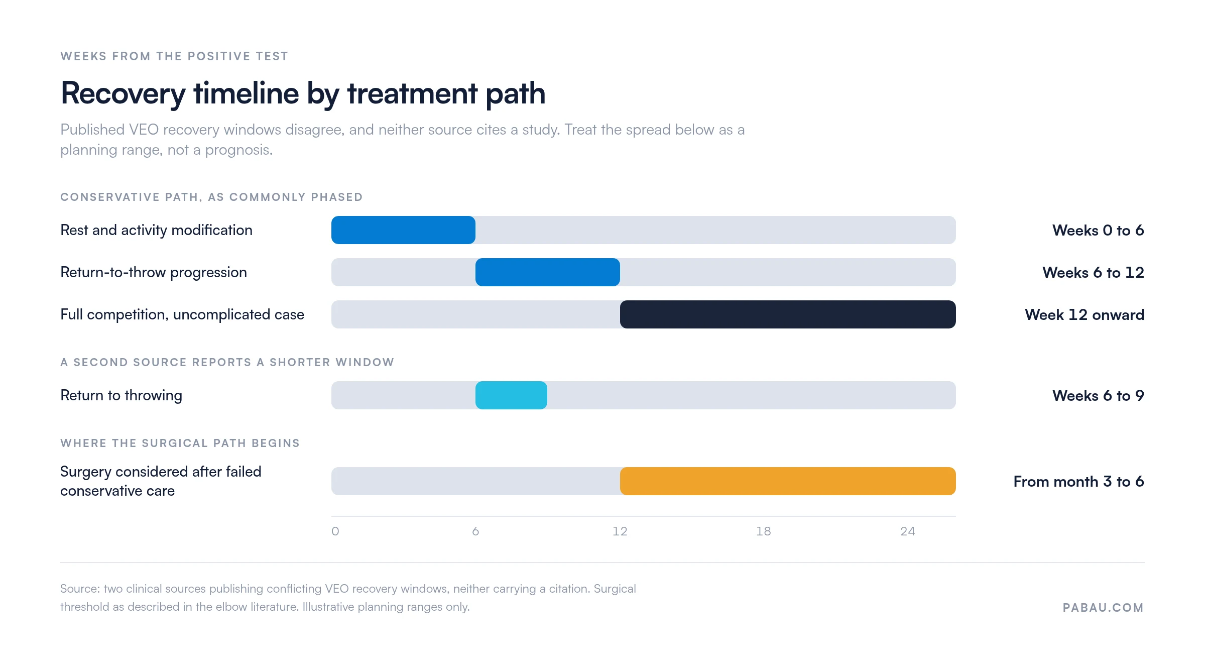
What drives the range sits outside both sources. Osteophyte size, whether a loose body is present, UCL status, and how much the athlete’s mechanics contributed all move the date. So does the competitive calendar.
The defensible position is to give the athlete a range and the criteria that close it. Full pain-free extension, restored flexor-pronator strength, and a completed interval throwing program are all checkable. A date on a calendar is not.
Phase criteria beat phase dates in any return-to-sport program, not only this one. The return to running protocol lays out the same criterion-based structure for a different population.
Rehab for VEO syndrome runs through five phases. You restore extension, rebuild the flexor-pronator group, stabilize the scapula, then correct the kinetic chain that loads the elbow. An interval throwing program closes it out.
Progression is criterion-based rather than time-based at every step. An elbow stiffness exercises handout gives you a patient-facing sheet for the extension work in phase one, which is the part athletes skip.
Anti-inflammatory medication and a selective corticosteroid injection can control acute symptoms. Neither replaces the load work above.
Treatment depends on symptom severity, imaging findings, UCL integrity, and the athlete’s competitive calendar. Most clinicians run a conservative-first pathway and reserve surgery for cases that do not settle.
Nonoperative management is first line for the majority of athletes with VEO syndrome. Its core components are:
Operative management is indicated when conservative care fails after an adequate trial, typically three to six months. It also applies when the osteophyte is large enough to cause mechanical symptoms regardless of rest, or when a loose body blocks extension.
The standard procedure is arthroscopic excision of the posteromedial olecranon osteophyte. Arthroscopy gives direct visualization of the posteromedial compartment, allows assessment of concurrent UCL pathology, and carries lower morbidity than an open approach.
Two technical points from the arthroscopy literature are worth knowing before you refer. Resecting the posteromedial osteophyte beyond its native margin, or by more than about 3 mm, raises stress on the medial collateral ligament. That over-resection can create instability the athlete did not have before.
A nick-and-spread technique near the medial portals protects the ulnar nerve during access. After resection, an arthroscopic valgus stress test checks for medial gapping that would indicate an incompetent ligament. Ulnar nerve decompression or transposition is sometimes done at the same time.
Post-operative timelines carry the same uncertainty as the nonoperative ones. The published windows conflict, so the honest answer to an athlete is a range with criteria attached.
VEO risk falls when throwing workload is capped and rest is enforced between outings. MLB and USA Baseball publish age-banded daily pitch limits through the Pitch Smart program, and those limits are the most concrete guidance available.
Rest requirements scale with the count thrown, not just the daily cap. A 13 or 14 year old who throws 66 to 95 pitches needs four days before pitching again. Figures come from the Pitch Smart guidelines published by MLB and USA Baseball.
The counting only helps if it covers the whole workload. Practical measures for a practice advising a throwing athlete:
None of this removes VEO risk from a career of overhead throwing. It moves the odds, and it gives you a workload record to point at when an athlete presents with posteromedial pain.
Sports medicine and physical therapy practices working with throwing athletes carry a specific documentation load. Each athlete needs repeated structured assessments over months, and the exam findings, imaging results, rehab milestones, and return-to-sport criteria all have to stay together.
Practice management software like Pabau keeps those pieces in one client record. You can build a throwing-athlete intake and examination template, so symptom onset, throwing volume, pain arc, and each test result get captured at every visit.
Outcome tracking is where the recovery framework above becomes usable. Recording extension range, flexor-pronator strength, and pain scores at each visit turns a vague timeline into criteria you can show the athlete. The same record holds pitch counts and rest days between outings.
For a multi-practitioner practice, that matters most at handover. Physiotherapy clinic management software centralizes the record, so whoever sees the athlete next picks up where the last clinician stopped.

Pabau keeps exam findings, imaging results, and rehab milestones in one client record. Sports medicine and physical therapy practices can follow an athlete through every phase without chasing notes.

The valgus extension overload test earns its place because it replicates the mechanism it is trying to find. Perform it properly, holding valgus stress through the arc from 70 degrees to full extension. A positive result then tells you where to look next.
What it cannot do is close the case. There is no accuracy figure for it, so imaging still decides. And the recovery conversation that follows is a range, not a date, whatever the published numbers imply.
The practical shift is small. Give the athlete criteria instead of a calendar, record the workload that got them here, and the return-to-sport decision stops being a guess.
Keeping that record consistent across a whole athlete roster is a systems problem, not a clinical one. Book a demo to see how Pabau structures exam findings and rehab tracking for sports medicine practices.

Need a criterion-based return-to-sport framework? Return to running protocol for physical therapy lays out a phased progression you can adapt for a throwing athlete.
Assessing the thrower’s shoulder too? O’Brien’s test covers the active compression maneuver for labral pathology, which often sits alongside elbow symptoms in overhead athletes.
Setting up or growing a musculoskeletal practice? Opening a physiotherapy clinic outlines the operational and compliance requirements that apply to a new physical therapy practice.
Screening the overhead athlete’s shoulder as well? Shoulder range of motion chart template gives you a printable chart for recording the rotation deficits that often sit behind elbow symptoms.
The valgus extension overload test is a clinical examination maneuver used to diagnose VEO syndrome in overhead throwing athletes. The examiner applies a valgus stress to the elbow while passively extending it from approximately 70 degrees of flexion to full extension. Reproduction of posteromedial elbow pain constitutes a positive result, indicating olecranon impingement against the posteromedial fossa.
Seat the patient with the affected arm relaxed. Stabilize the humerus with one hand, then apply a valgus force at the wrist or distal forearm with the other. While maintaining that valgus load, passively extend the elbow from roughly 70 degrees to full extension. Ask the patient to report any pain during the arc, and note its location and the point in the arc where it appears.
No published sensitivity or specificity value exists for the VEO test. No prospective study has validated it against arthroscopic findings as a reference standard. The test is used empirically because it directly replicates the throwing mechanism. Clinicians treat a positive result as a strong indicator warranting imaging confirmation rather than a standalone diagnostic finding.
The VEO test targets posteromedial impingement and osteophyte pathology by extending the elbow from 70 degrees to full extension under valgus stress. The moving valgus stress test targets UCL integrity by applying valgus stress through the 70 to 120 degree arc. That arc is the shear stress zone for the UCL during pitching. The MVST has stronger published accuracy data for UCL pathology. Both are typically used together when examining a throwing athlete.
Yes. VEO syndrome is most common in overhead throwing athletes, such as baseball pitchers and cricket bowlers. Any activity combining repetitive valgus stress with rapid elbow extension can produce the same pathology. Gymnasts, offensive linemen, and racquet sport athletes occasionally present with VEO syndrome, though at lower prevalence than throwing athletes.
Plain radiographs, particularly posteromedial oblique views, are the first-line imaging modality for detecting posteromedial osteophytes. MRI is preferred when concurrent UCL pathology or bone edema is suspected. CT is used selectively for preoperative planning, to map osteophyte morphology and identify loose bodies more precisely than a radiograph or MRI can.
No. ICD-10-CM has no dedicated code for valgus extension overload syndrome as a named entity. M21.021 covers valgus deformity of the right elbow, but that describes a structural deformity rather than this overuse impingement syndrome. In practice, clinicians code the specific finding documented, such as an olecranon osteophyte, posterior elbow impingement, or an elbow sprain. Confirm the current-year code with your billing or coding reference before you submit.