3 Hour Glucose Test Results Chart
A 3 hour glucose test results chart lists the cutoff for each blood draw in the ...

1
Last Updated: Oct 14, 2026

Snapping hip syndrome (coxa saltans) has three subtypes: external (IT band), internal (iliopsoas), and intra-articular. Each one calls for a different special test.
The snapping hip test is not a single maneuver. Clinicians combine the Ober test, the internal snapping hip maneuver, FABER, FADIR, and active provocation to localize the structure.
Internal snapping hip is confirmed by guiding the hip from flexion, abduction, and external rotation into extension. An anterior snap during that extension is the positive sign.
The Thomas test measures hip flexor length only. A tight iliopsoas supports the diagnosis as a contributing factor, but the test carries no snap sign of its own.
Dynamic ultrasonography is the gold-standard imaging adjunct for tendinous snapping hip. When intra-articular pathology such as a labral tear is suspected, MRI or MR arthrography is preferred.
Pabau’s digital intake forms and client records help physical therapy and MSK practices document hip assessments, track rehabilitation progress, and automate follow-up.
A clicking hip is one of the most under-documented findings in MSK practice. Most patients have lived with it for months before mentioning it, often because it is painless. Pain changes that. Once the snap hurts, or once it interferes with sport, the clinical task changes. Identify the structure, confirm the subtype, and rule out intra-articular pathology. The snapping hip test is where that process begins.
This guide is written for physical therapists, sports medicine clinicians, and MSK practitioners. It covers how to perform and interpret the key special tests for snapping hip syndrome. It also covers the anatomy behind each subtype, imaging adjuncts, differential diagnosis, and rehabilitation.
The snapping hip test is not a single maneuver. It refers to a structured battery of clinical examination tools. Together they identify which structure is causing the audible or palpable snap around the hip. Snapping hip syndrome (SHS), also called coxa saltans, is classified by the location of the offending structure.
The three subtypes rarely present identically. Location of the snap, associated pain, and aggravating activities all guide which tests to prioritize.
The anatomy explains what each test is provoking, and every subtype has its own biomechanical account.
External SHS: The iliotibial band (IT band) or the posterior border of the gluteus maximus passes over the prominent greater trochanter. That happens during hip flexion and extension. When the band is tight or the trochanter is particularly prominent, it catches and releases with a palpable, sometimes audible, snap on the lateral hip. This is the most common subtype and frequently seen in runners and cyclists.
Internal SHS: The iliopsoas tendon snaps over the iliopectineal eminence or the anterior femoral head. That eminence is the bony ridge at the junction of the ilium and pubis. The snap arrives during hip flexion, abduction, and external rotation. Patients typically describe an anterior groin snap, sometimes with clicking and groin pain. Stair climbing and rising from a seated position are the usual triggers.
Intra-articular SHS: Rather than a tendinous mechanism, the snap originates within the joint itself. Loose bodies, acetabular labral tears, or osteochondral defects cause mechanical clicking that is hard to reproduce consistently on examination. It usually arrives with joint-line pain, locking, or giving way.
Each named test below targets a specific subtype. Perform them in sequence to build a complete clinical picture before considering imaging.
The Ober test evaluates IT band tightness, the primary driver of external SHS. A positive test in the context of a laterally located snap is strongly suggestive of coxa saltans externa.
Knee position changes what the test loads, so technique varies between examiners. The dedicated guide to the Ober test covers the modified version and the reliability data behind each variant.
This is the maneuver that reproduces the iliopsoas snap, and it is what confirms internal SHS. It takes the hip out of flexion, abduction, and external rotation, then carries it into extension. That is the arc in which the tendon crosses the iliopectineal eminence and catches.
The direction of travel matters. The snap belongs to the extension phase, not to the flexion phase and not to a static end position. A hip that clicks only at end-range flexion should be taken through FABER and FADIR testing instead.
The Thomas test measures hip flexor length. It is a static length test with no snap sign, so it cannot confirm internal SHS on its own. Use it to find the shortened iliopsoas that often sits behind a snapping tendon. It also sets a baseline you can retest after a stretching program.
Read a positive Thomas test as a contributing factor, not as a diagnosis. Plenty of patients with short hip flexors never snap, and plenty of snapping hips lower flat to the table. Confirmation still comes from the FABER-to-extension maneuver above.
That baseline is only useful if the angles are recorded the same way at every review. A printable Thomas test template gives you the fields for thigh and knee position, so the retest is comparable months later.
FABER stands for flexion, abduction, and external rotation. This test screens for intra-articular hip pathology, including labral tears and sacroiliac joint dysfunction, helping to differentiate them from tendinous snapping.
FADIR (flexion, adduction, internal rotation) is the primary provocation test for femoroacetabular impingement (FAI) and labral pathology. It complements the FABER test when intra-articular SHS is suspected.
Circumduction is the maneuver to reach for when the named tests come back equivocal. It lets the patient move through the whole arc under load, which often reproduces a snap that passive testing misses.
Individual test results only help when they are read together. A positive Ober test is sometimes taken as proof of bursitis rather than IT band tightness. A positive FADIR is easy to overlook in a patient who reports a click rather than pain. The table below maps each result to its most likely subtype and the correct clinical next step.
One caveat applies to all of these maneuvers. Reproducing the snap identifies the structure, but it does not rule out a second problem inside the joint. Dynamic ultrasonography is the gold-standard confirmation tool for tendinous snapping, as reported in the orthopedic literature indexed in PubMed Central. Where the findings point inside the joint instead, MRI or MR arthrography is the study to request.
Several hip conditions share overlapping presentations with snapping hip syndrome. Missing a differential can delay appropriate management by months.
Several of these differentials separate more cleanly on a full regional screen than on any single provocation test. A structured hip examination adds range of motion, gait, and neurovascular findings to the special tests above. When the picture is still unclear after that, imaging is the appropriate next step.
Clinical tests alone may be insufficient when the presentation is atypical or intra-articular pathology is suspected. Which study to request follows from what the examination provoked, and the two routes diverge sharply.
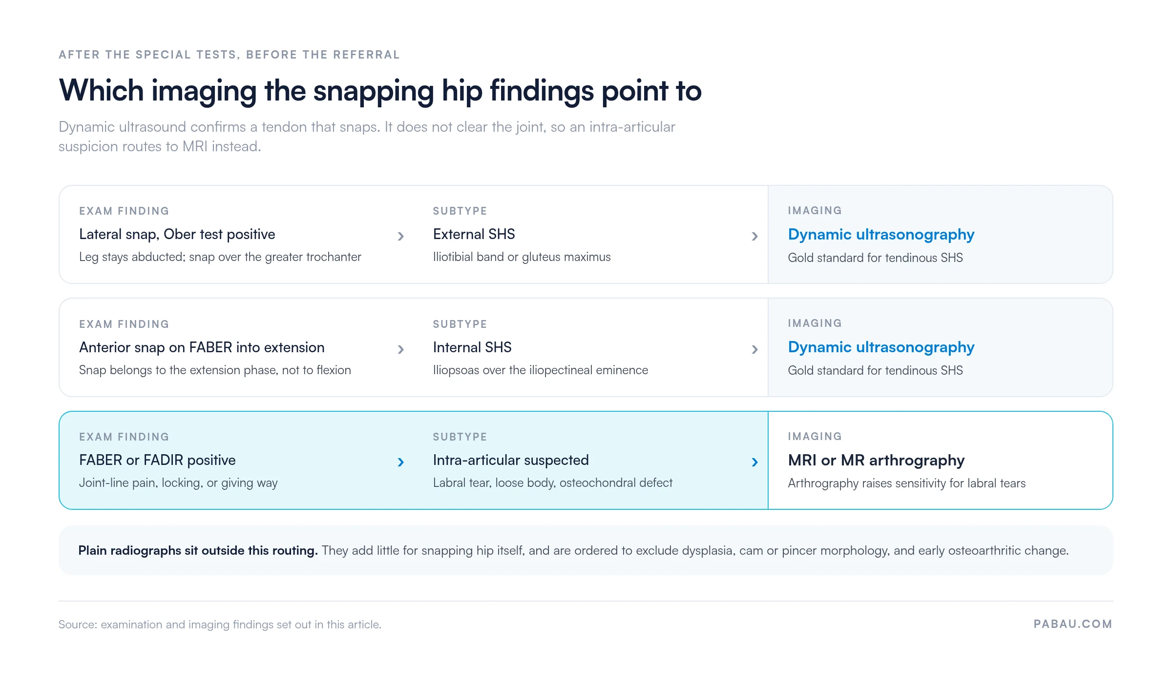
Dynamic ultrasound should be considered the first-line imaging tool for tendinous SHS. Refer to MRI when FABER or FADIR testing raises concern for labral or intra-articular pathology.
The majority of snapping hip syndrome cases respond to conservative management. Surgical referral is rarely needed in the absence of intra-articular pathology.
Treatment targets IT band flexibility and lateral hip strength. A weak gluteus medius allows excessive hip adduction during running and stair descent, increasing IT band tension over the greater trochanter.
Rehabilitating an athlete after external SHS means staging the return to load. A structured return-to-running protocol takes the progression from pain-free walking through to full training.
Iliopsoas stretching reduces the tendon tension that drives snapping over the iliopectineal eminence. Core strengthening improves lumbopelvic control, reducing the compensatory anterior pelvic tilt that places sustained load on the hip flexors.
Certain exercises reproduce or aggravate the snapping mechanism and should be avoided during the acute and sub-acute phases.
Document the exact location, quality (audible vs. palpable only), and aggravating activities for every snapping hip presentation. That baseline lets you track rehabilitation progress objectively. It also flags the cases where the snap worsens or turns painful, which is your signal to escalate to imaging.
Most patients with snapping hip syndrome do not require surgical intervention. Refer when the following criteria are met after 3 to 6 months of structured conservative management.
Surgical options for refractory cases include iliopsoas tendon lengthening (for internal SHS) and IT band Z-plasty or release (for external SHS). Arthroscopic labral repair or debridement addresses intra-articular causes.
The Orthobullets clinical reference on coxa saltans reports that conservative management resolves symptoms in most patients. Surgical referral is reserved for those who fail non-operative treatment. Outcomes data from Orthopedic Reviews supports return to sport in most patients after conservative or surgical management.
Accurate assessment is only half the job. The practice still has to record which tests were performed, what each one provoked, and when the patient is due back. Practice management software like Pabau is built for that kind of multi-stage patient journey.
Clinicians can capture structured hip assessment data on a digital intake form before the patient arrives. The findings then sit in a timestamped client record that carries test results forward across appointments. Automated workflows send rehabilitation reminders and recall patients at the six-week and three-month marks, which matches the conservative timeline above.
Pabau is software for physical therapists and the wider MSK team, so notes, scheduling, and recall sit in one record rather than three systems. That leaves you a rehabilitation history you can audit at any review point, without reconstructing it from memory.


Pabau helps physical therapy and sports medicine practices document hip assessments, manage follow-up care, and automate patient recall so no rehabilitation milestone gets missed.

The judgment worth getting right comes at the end of the examination, not the start. A snap you can reproduce on the table is a tendon, and it routes to dynamic ultrasound. A hip that hurts on FABER or FADIR without a reproducible snap belongs on the MRI pathway, whatever the patient calls the noise.
Painless snapping needs documentation and reassurance more often than it needs a scan. Painful or worsening snapping earns a defined conservative block of three to six months. Set the retest date at the first appointment, so the referral decision is made against a baseline rather than a recollection.
Pabau’s documentation tools help MSK and physical therapy teams capture assessment findings and automate recall at each rehabilitation milestone. To see how that works across a full patient journey, book a demo with our team.

Need to tell a strain from a snap? Hip flexor strain test sets out the resisted-load and palpation findings that point to a torn muscle rather than a snapping tendon.
Checking whether the iliopsoas is genuinely short? Tight hip flexors test covers the length testing and the retest points that show whether a stretching program is working.
Tracking hip function across a rehabilitation block? Harris Hip Score gives you a scored outcome measure to record at baseline and at every review.
Managing athlete rehabilitation timelines? Return-to-running protocol for physical therapy provides a progressive load framework for guiding athletes from protected weight-bearing through to full training.
The snapping hip test is a clinical examination battery used to find the cause of snapping or clicking around the hip. It is not a single maneuver. Clinicians combine the Ober test for the IT band with the FABER and FADIR tests for intra-articular pathology. Internal snapping hip is confirmed separately, by guiding the hip from flexion, abduction, and external rotation into extension.
Snapping hip syndrome is caused by a tendon or band catching on a bony prominence during hip movement. The most common cause is the iliotibial band snapping over the greater trochanter, which is the external type. The other is the iliopsoas tendon snapping over the iliopectineal eminence, the internal type. Intra-articular causes include labral tears, loose bodies, and osteochondral defects within the hip joint itself.
External snapping hip involves the iliotibial band or gluteus maximus snapping over the greater trochanter on the lateral side of the hip. Internal snapping hip involves the iliopsoas tendon catching on the iliopectineal eminence or femoral head at the anterior groin. The location of the snap, the associated symptoms, and which special tests reproduce the click are the key distinguishing features.
See a clinician when the hip click is painful, getting louder, or getting more frequent. The same applies when it comes with groin pain or locking, or interferes with sport and daily activity. Painless clicking in isolation that has been present for years without change is low priority. A sudden onset of painful clicking following trauma or a fall warrants prompt assessment to exclude hip labral injury or fracture.