



GLP-1 diet plan
A GLP-1 diet plan is a structured eating framework for patients taking semagluti...

2
Last Updated: Oct 13, 2026

Kleiger’s test (the external rotation stress test) screens for syndesmosis and deltoid ligament injury, not lateral ligament sprains
Positive sign: anterolateral pain indicates syndesmosis involvement; medial pain indicates deltoid ligament disruption
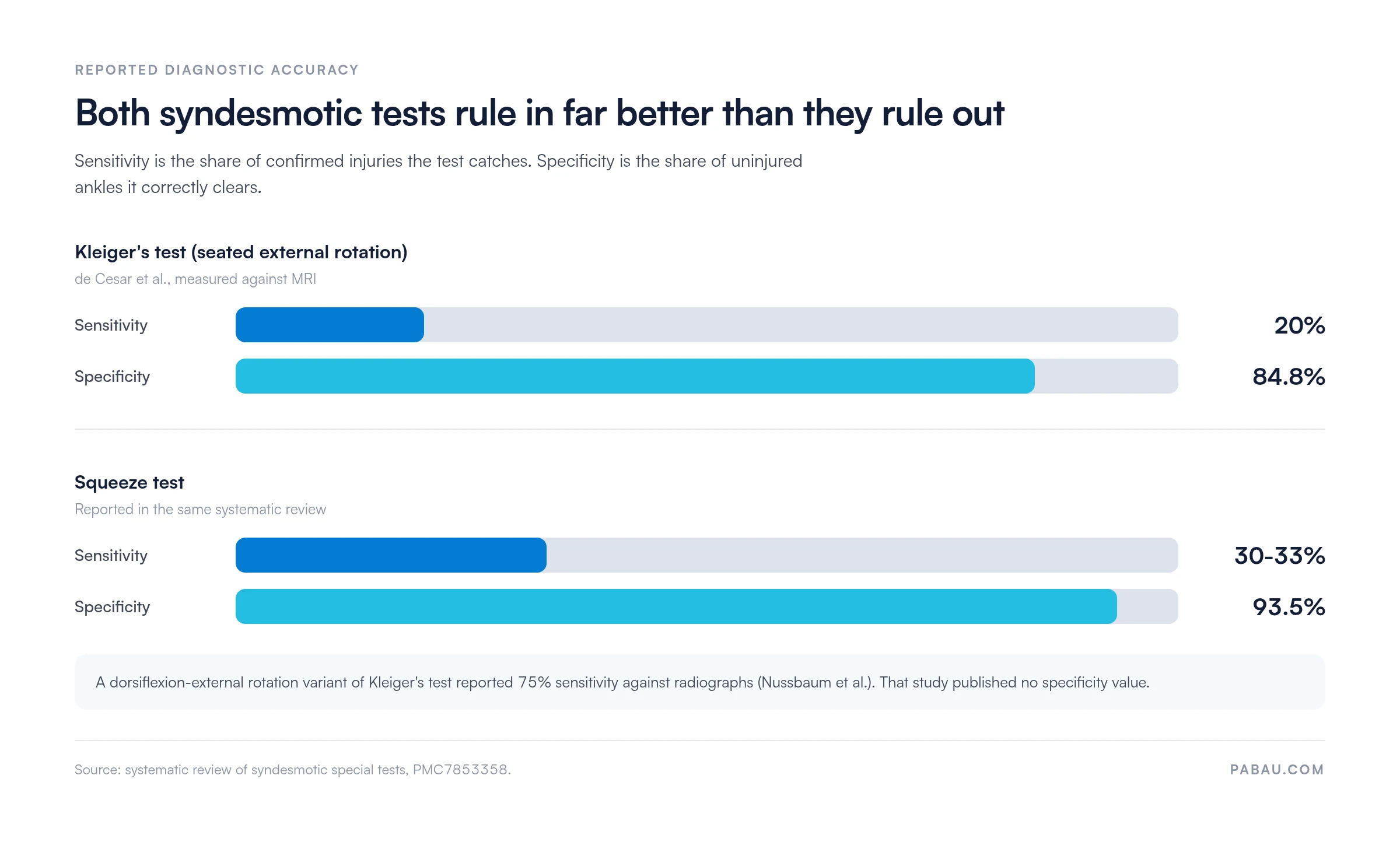
Reported sensitivity is low (20% against MRI) and specificity is 84.8%, so a positive finding rules in better than a negative rules out
No published study has established the test’s accuracy for deltoid ligament injury specifically
Pabau’s digital forms and clinical records let physiotherapy and sports medicine teams document findings immediately at the point of care
Kleiger’s test is a clinical orthopedic special test used to detect ankle syndesmosis injury (high ankle sprain) and deltoid ligament disruption.
The examiner applies an external rotation force to the foot while the patient sits with the knee flexed to 90 degrees. It is one of the most widely used syndesmotic special tests.
Pain over the anterolateral ankle points to the syndesmosis, while medial pain points to the deltoid ligament. That distinction shapes the imaging referral and the return-to-sport timeline. Reported sensitivity is low at 20% against MRI, and specificity is 84.8%, so a positive finding carries more weight than a negative one.
This guide covers the anatomy, the step-by-step technique, result interpretation, diagnostic accuracy, and the clinical next steps that follow a positive finding.
Kleiger’s test is an ankle orthopedic special test that stresses the distal tibiofibular syndesmosis and the medial deltoid ligament. It does so through a controlled external rotation force. It is also called the external rotation stress test, and the two names are interchangeable across clinical literature. Physical therapists, sports medicine physicians, and orthopedic clinicians typically reach for it when the injury mechanism suggests a syndesmotic rather than a lateral ligament sprain.
Two distinct injury patterns make Kleiger’s test the right choice. First, a high ankle sprain caused by external rotation or hyperdorsiflexion forces. Those forces drive the talus outward against the fibula, stressing the anterior inferior tibiofibular ligament (AITFL) and the wider syndesmotic complex. Second, medial ankle pain following a forced external rotation or eversion mechanism, which may indicate deltoid ligament disruption. A lateral ligament sprain from inversion, by contrast, would not stress these structures and the test would likely remain negative.
The test is indicated when the patient reports pain proximal to the lateral malleolus, or when they describe a mechanism involving external rotation. It is also indicated when the Ottawa ankle rules calculator flags the need for further structural evaluation. Physical therapists and sports medicine clinicians run it alongside weight-bearing assessment. Recording the finding in software for physical therapy gives the reassessment something to compare against.
The ankle syndesmosis is a fibrous joint connecting the distal tibia and fibula. It relies on four ligaments and a sheet of connective tissue to maintain the mortise width that allows the talus to fit securely during loading. Understanding these structures clarifies why external rotation produces the pain pattern Kleiger’s test exploits.
When external rotation force is applied to the foot, the talus acts as a wedge, pushing the fibula laterally and stressing the AITFL first. If the force continues, load transfers through the IOM toward the PITFL. Concurrent medial pain reflects tensile stress on the deltoid ligament as the talus translates. This anatomy is why a single test can screen for two structurally distinct injury patterns depending on where the patient localizes their pain.
The Kleiger’s test procedure requires precise patient positioning and a consistent examiner technique. Both are needed to reproduce the mechanism reliably and to avoid false positives from unrelated ankle instability.
Good documentation at this step matters. Clinicians using digital intake forms can pre-build an ankle examination template for the bedside. It captures pain location, provocation grade, and contralateral comparison, which reduces the risk of incomplete records before imaging referral. Clear note-writing matters most when the findings may influence a surgical versus conservative management decision.

A positive Kleiger’s test is pain reproduced over the syndesmosis or medial ankle during the external rotation maneuver. The location of that pain indicates which structure is involved.
The two distinct positive patterns carry different clinical implications:
A negative test does not rule out a syndesmotic injury. That holds particularly in a subacute or chronic presentation, where the acute inflammatory pain has already subsided. Clinical context, mechanism, and additional testing remain essential.
Kleiger’s test sensitivity is low and its specificity is moderate to high, so a positive result carries far more weight than a negative one. The systematic review indexed as PMC7853358 collects the diagnostic accuracy studies for syndesmotic special tests. Two of the included studies report figures for the external rotation test, and they used different maneuvers and different reference standards.
The two figures are not in conflict, because the studies did not measure the same thing. de Cesar et al. tested the seated external rotation maneuver against MRI. At 20% sensitivity, it missed four out of five confirmed syndesmotic injuries in that sample. Nussbaum et al. tested a dorsiflexion-external rotation variant against radiographs and reported 75% sensitivity, but published no specificity value.
What the numbers support in practice is narrow. Specificity of 84.8% means a clearly localized positive meaningfully raises the probability of syndesmotic involvement. Sensitivity that low means a negative test barely narrows the differential, particularly in a guarded or subacute presentation. Kleiger’s test belongs in a multi-test assessment, never as a standalone diagnostic tool.
The review states a second limit plainly. No study it identified established the accuracy of the external rotation test for detecting deltoid ligament injury specifically. The medial pain pattern described above rests on anatomical reasoning and clinical convention, not on a measured sensitivity or specificity. Treat a medial positive as a prompt for imaging rather than as evidence of deltoid disruption.
No single ankle syndesmosis test has sufficient diagnostic accuracy to stand alone. Combining Kleiger’s test with the squeeze test and fibula translation test improves overall diagnostic confidence for high ankle sprain.
Clinically, two positives point more firmly toward syndesmotic injury than one. The squeeze test carries its own low sensitivity in the same review, 30-33%, alongside specificity of 93.5%. Both tests therefore rule in better than they rule out, which is the reason to run them together rather than to pick between them.
Cadaveric work cited in the review offers one mechanical reason to keep Kleiger’s test in the sequence. External rotation produced more tibiofibular displacement than the squeeze, fibular translation, and Cotton tests. Running the pair takes under two minutes at the bedside.
A positive Kleiger’s test confirms syndesmosis involvement but does not grade severity. Grading determines whether the injury can be managed conservatively or requires surgical stabilization. The most widely referenced clinical classification divides syndesmotic sprains into three grades based on anatomical disruption.
Grade III injuries presenting with diastasis on a weight-bearing radiograph require orthopedic referral. Document the grading findings clearly, because the management decision made here sets the recovery timeline. That ranges from six weeks for Grade I to four to six months for a surgically managed Grade III injury. Knowing the grade also shapes return-to-sport planning, and the return-to-running protocol sets out post-clearance progression for lower-limb injuries.
Kleiger’s test has documented limitations that clinicians should factor into their assessment decision-making. These fall into four categories: patient factors, examiner factors, timing of assessment, and structural confounders.
Patients with high pain tolerance may not report pain even with significant ligamentous disruption. Conversely, patients with acute swelling and guarding may report pain with any movement, increasing the risk of false positives. Pain apprehension alone, without anatomically localized provocation, should not be recorded as a positive test.
Technique changes the result. Excessive force can provoke pain in an uninjured ankle, and letting the knee drift out of 90 degrees alters the torque reaching the mortise. Grip pressure over the distal tibia and fibula can reproduce syndesmotic pain on its own, which reads as a false positive. Standardizing hand position and rate of rotation across a team keeps serial reassessments comparable.
Acute injuries with significant edema reduce the specificity of pain localization. The patient may report diffuse ankle pain rather than pinpointing the syndesmosis or deltoid. Reassessment at 48-72 hours after initial swelling reduction often yields more reliable findings. Subacute and chronic syndesmotic sprains may produce minimal pain provocation even with incomplete healing, making the test less sensitive in this phase.
Medial ankle pain during external rotation does not exclusively indicate deltoid ligament injury. Tibialis posterior tendinopathy, medial talar dome chondral lesions, and medial gutter impingement can all produce medial pain with this maneuver. Similarly, anterolateral pain may reflect anterior ankle impingement or a distal fibular stress reaction rather than syndesmotic disruption. These differential diagnoses require imaging to exclude.
Structured assessment protocols help a busy caseload. They flag these differentials at the documentation stage and prompt consistent referral pathways. Teams who track outcomes against imaging findings over time can build local sensitivity data for their own patient population. Most physiotherapy practice management software will hold that outcome data alongside the original examination note.

When assessing for a high ankle sprain in the acute phase, perform the squeeze test before Kleiger’s test. Proximal compression is less likely to provoke guarding than distal rotation, giving you a cleaner first provocation signal. Reserve Kleiger’s as your confirmatory second step.
A positive Kleiger’s test opens a structured assessment pathway. Which step comes next depends on the suspected grade of injury and on whether plain films show a bony abnormality.
Documenting the assessment pathway end to end supports physiotherapy compliance requirements. It covers the initial clinical findings, the imaging results, and the management decision. It also leaves an auditable clinical record if outcomes are questioned later. Embedding a syndesmosis template directly into the consultation workflow reduces reliance on paper, which is easily lost between referral stages.
A syndesmosis assessment produces findings that matter weeks later. Pain location, suspected grade, the imaging decision, and the reassessment date all feed into return-to-sport clearance. In most practices that trail is split across a paper assessment sheet, a scanned radiology report, and a note typed up later. By the time a colleague covers the follow-up, half of it has to be reconstructed from memory.
Practice management software like Pabau keeps that chain in one patient record. You can build an ankle examination form that captures pain site, provocation grade, and contralateral comparison as structured fields. Imaging results, referral letters, and reassessment notes attach to the same timeline. The record no longer travels on paper between the treatment room and the filing cabinet.
The result is a record a covering clinician can read without calling you first. It also gives the practice an auditable account of why a conservative or surgical pathway was chosen, should that decision be questioned later.

Pabau’s digital forms and client records let physiotherapy and sports medicine teams capture examination findings, referral decisions, and treatment plans at the point of care. Admin drops, and each patient’s clinical history stays in one place.

Run Kleiger’s test for what it localizes, not for what it confirms. A clearly anterolateral positive raises the probability of syndesmotic involvement enough to justify weight-bearing films. A negative result changes very little, so let the mechanism and the swelling pattern keep driving the decision.
The medial finding is the trade-off worth remembering. No published study has measured the test’s accuracy there. Treat medial pain as a reason to image rather than as a deltoid diagnosis. Pair the test with the squeeze test, reassess at 48 to 72 hours, and grade the injury before committing to a management pathway.
Standardizing that sequence across a team is mostly a documentation problem. Book a demo to see how Pabau keeps ankle examination findings, imaging results, and reassessment notes in one patient record.

Assessing the Achilles in the same visit? Thompson test covers the calf-squeeze screen for Achilles tendon rupture and how to interpret it.
Want a second decision rule before ordering films? Bernese ankle rules gives you a printable indirect-stress alternative to the Ottawa criteria.
Recording radiograph findings after referral? Ankle radiograph results template structures clear space, overlap, and mortise measurements in one sheet.
Need to rule out posterior talofibular involvement? Ankle posterior drawer test template walks through the technique and the recording fields.
Need a framework for ankle assessment compliance? Mandatory compliance for physiotherapy clinics covers the documentation and audit requirements for musculoskeletal practice.
Kleiger’s test screens for two conditions: ankle syndesmosis injury (high ankle sprain, involving the AITFL, IOM, and/or PITFL) and deltoid ligament disruption. It does not assess the lateral ligament complex, so it is not appropriate for standard inversion ankle sprains involving the ATFL or CFL.
A positive Kleiger’s test is pain reproduced during controlled external rotation of the foot. Anterolateral pain indicates syndesmotic involvement, and medial pain indicates deltoid ligament involvement. Visible lateral translation of the talus within the mortise is an additional positive finding indicating significant instability.
Kleiger’s test stresses the syndesmosis through a rotational mechanism at the ankle. The squeeze test compresses the tibia and fibula proximal to the syndesmosis, generating indirect stress distally. They provoke pain through different mechanisms and complement each other. A positive finding on both increases diagnostic confidence substantially compared to either test alone.
The PMC7853358 systematic review reports 20% sensitivity and 84.8% specificity for the seated external rotation test, measured against MRI. A second study in the same review reported 75% sensitivity for a dorsiflexion-external rotation variant against radiographs, with no specificity value published. No study in the review established the test’s accuracy for deltoid ligament injury specifically. These figures support using Kleiger’s test as part of a multi-test assessment rather than in isolation.
It can suggest one, but it cannot confirm one. Medial ankle pain provoked during external rotation indicates tensile stress on the deltoid ligament complex. No published study has measured the test’s accuracy for deltoid injury. Medial pain can also arise from tibialis posterior tendinopathy or medial talar dome pathology. Imaging is needed before management decisions are made.
Order a weight-bearing AP and mortise-view radiograph first. A tibiofibular clear space above 6 mm indicates diastasis and warrants urgent orthopedic referral. If radiographs are normal but clinical suspicion remains high, MRI is the reference standard for soft tissue assessment. Grade I-II injuries follow conservative rehabilitation, and Grade III injuries with diastasis are referred for surgical stabilization.






