



GLP-1 diet plan
A GLP-1 diet plan is a structured eating framework for patients taking semagluti...

2
Last Updated: Oct 13, 2026

The cerebellar examination is a structured clinical assessment evaluating coordination, balance, gait, and motor control through systematic physical tests.
The DANISH mnemonic (Dysdiadochokinesia, Ataxia, Nystagmus, Intention tremor, Slurred speech, Hypotonia) summarizes the six cardinal cerebellar signs to identify dysfunction.
Common causes of abnormal findings include stroke, multiple sclerosis, alcohol excess, space-occupying lesions, and hereditary ataxias. Accurate documentation supports clinical decision-making.
Using a downloadable cerebellar examination checklist streamlines OSCE preparation, standardizes clinical documentation, and ensures consistency across healthcare teams.
A ready-to-use structured template for performing and documenting the cerebellar examination, covering gait assessment, coordination testing, eye movements, speech evaluation, and standardized interpretation of findings.
Download templateClinicians performing neurological exams need a reliable structure to assess cerebellar function consistently. The cerebellar examination is a cornerstone of neurological assessment in physical therapy practice and beyond, yet many students and junior practitioners find the sequence of tests and interpretation of findings unclear. This guide walks you through each step of the cerebellar examination with clinical context and documentation guidance.
Whether you’re preparing for an OSCE station, managing patients with ataxia or balance disorders.
The cerebellar examination is a systematic clinical assessment designed to evaluate the function of the cerebellum and detect signs of cerebellar dysfunction. It tests coordination, balance, motor control, gait, speech, and eye movements, all functions the cerebellum governs.
Healthcare professionals use this examination to identify patients with ataxia, intention tremor, dysarthria, or other cerebellar signs that indicate disease in the posterior fossa, brainstem, or cerebellum itself.
The exam is a standard component of the full neurological assessment and is particularly important in evaluating patients with suspected stroke, multiple sclerosis, alcohol-related cerebellar degeneration, tumors, or hereditary ataxias. Unlike other neurological examinations, the cerebellar exam requires the patient to be upright and mobile for much of the testing, so environment and patient cooperation are critical.
A structured neurological assessment template ensures that each component of the cerebellar examination is performed in a logical order, building from observation to specific testing. Follow this sequence to conduct a thorough examination.
Before any formal testing, observe the patient’s appearance, posture, and movement. Note whether they use walking aids, whether they have tremor at rest, and whether their balance appears affected even when seated.
Ask the patient to speak naturally, then specifically ask them to repeat phrases. Listen for scanning speech (characteristic of cerebellar dysfunction) or slurred speech that worsens with fatigue.
Test smooth pursuit, saccadic eye movements, and assess for gaze-evoked nystagmus. Cerebellar lesions typically produce horizontal nystagmus that is worse when looking toward the side of the lesion.
These tests reveal intention tremor, dysmetria, and dysdiadochokinesia, all hallmarks of cerebellar pathology.
The heel-shin test is the key lower limb assessment. The patient lies supine and runs their heel down the opposite shin.
Observe the patient walking along a line. Cerebellar ataxia produces a broad-based, unsteady gait.
The patient stands with feet together, eyes first open then closed. A positive Romberg test (falls or sways significantly with eyes closed) suggests proprioceptive loss, NOT cerebellar dysfunction. Cerebellar ataxia causes difficulty even with eyes open.
During your examination, you’ll be looking for the cardinal cerebellar signs summarized by the DANISH mnemonic. This quick reference helps you remember all six key findings and link them to patient assessment workflows.
Teams using software to manage patient records can pull up prior scores before every reassessment.
A downloadable cerebellar examination checklist streamlines the assessment process. Use this template during clinical practice, OSCE preparation, or team training.
The cerebellar examination produces findings that must be interpreted in clinical context. Multiple signs from the DANISH framework, especially in combination, point toward cerebellar dysfunction rather than other neurological disorders. The pattern of findings can also suggest the location of the cerebellar lesion.
A positive finding in even one DANISH sign warrants further investigation. For example, isolated intention tremor might indicate early cerebellar disease or a reversible cause like medication toxicity, whereas a full constellation of DANISH signs (ataxia, dysarthria, nystagmus, dysdiadochokinesia, hypotonia) indicates significant cerebellar pathology.
When a patient presents with abnormal cerebellar examination findings, consider these etiologies:
Proper documenting clinical examination findings in your records ensures the clinical team can track progression and inform referral decisions.
Record cerebellar examination findings with precision. Document whether each DANISH component is present, absent, or not assessed. Include specific observations, not just interpretations.
Use comprehensive client records that support structured clinical documentation. This ensures your cerebellar examination findings are integrated into the full neurological assessment and available to your entire team. General practice generates results that arrive after the appointment, so the record has to bring them back together.
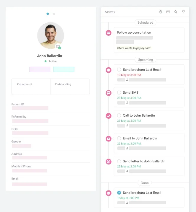

Digital forms and structured templates from practice management software like Pabau help you document neurological assessments consistently across your practice.

The structured cerebellar examination you practice for OSCE is directly applicable in real clinical settings. The same systematic approach that earns marks in an exam also ensures you don’t miss findings in a patient presenting with dizziness, ataxia, or suspected stroke.
In practice, you may need to adapt the examination. An acutely unwell patient may not be able to stand for full gait testing, so you perform what you can and document it honestly. The framework still applies: inspection, speech, eyes, upper limb, lower limb, gait, Romberg.
The key is building muscle memory so the sequence becomes automatic. When you’re assessing a real patient in a busy practice, automated clinical workflows help you complete each component without rushing. This clinical rigor is what distinguishes a competent examination from a cursory one.
A structured cerebellar examination is one of the highest-yield components of the full neurological assessment. By following a systematic approach and using the DANISH mnemonic to anchor your findings, you’ll reliably detect cerebellar dysfunction and guide your clinical team toward the correct diagnosis.
The downloadable template here supports both OSCE preparation and everyday clinical practice. Book a demo to see how Pabau’s clinical assessment tools help your team document and track neurological examinations consistently across your practice.

Assessing a patient with burn injuries? burn percentage chart calculates total body surface area affected by burns to guide treatment and fluid resuscitation.
Helping a family plan a move into assisted living? assisted living move-in checklist walks through the pre-move-in, move-in, and after-move-in tasks a family needs to track.
Caring for a patient with cholecystitis? cholecystitis nursing care plan template structures assessment triggers, intervention rationales, and discharge planning for acute and chronic cases.
DANISH stands for the six cardinal cerebellar signs: Dysdiadochokinesia (inability to perform rapid alternating movements), Ataxia (incoordination and unsteady gait), Nystagmus (involuntary eye movements), Intention tremor (tremor worsening as limb approaches target), Slurred speech or dysarthria (scanning or explosive speech), and Hypotonia (decreased muscle tone).
The Romberg test specifically identifies proprioceptive loss. A positive Romberg (significant swaying or falling only when eyes are closed) indicates loss of position sense, NOT cerebellar dysfunction. Cerebellar ataxia causes difficulty standing and walking even with eyes open because the cerebellum coordinates movement regardless of visual input.
The patient lies supine and places their heel on the opposite knee, then slowly slides the heel down the shin toward the ankle. Dysmetria (the heel deviates off the midline) or intention tremor (visible wavering) indicates cerebellar dysfunction. Perform bilaterally and compare sides.
The most common causes are posterior circulation stroke, multiple sclerosis, chronic alcohol excess, space-occupying lesions (tumors or cysts), and hereditary ataxias. Less common causes include drug toxicity (anticonvulsants, chemotherapy), infection, and metabolic disorders. Acute sudden-onset cerebellar signs warrant urgent imaging.
Yes. This guide provides access to a downloadable Cerebellar Examination Template that you can use for OSCE preparation, clinical practice, or team training. The template covers all examination steps, the DANISH mnemonic, and space for recording findings and clinical interpretation.
Ask the patient to perform rapid pronation-supination (flipping forearms in and out) or rapid toe tapping. Cerebellar dysfunction produces clumsy, irregular, uncontrolled movements because the cerebellum cannot coordinate the alternating agonist-antagonist muscle activation needed for smooth alternating movement. It specifically indicates cerebellar pathology.






