Nitrates medication list
A nitrates medication list is a point-of-care reference that sets out the main c...

Last Updated: Oct 6, 2026
The Royal London Hospital test is a bedside palpation test for mid-portion Achilles tendinopathy. The examiner finds the most tender point on the tendon with the ankle relaxed in plantarflexion. That same point is then palpated again with the ankle held in maximal dorsiflexion. The test is positive when the tenderness disappears or drops sharply in dorsiflexion.
The arc sign is a different test, and the two are often confused. It is positive when the tender, swollen segment of tendon moves with the tendon as the ankle is dorsiflexed and plantarflexed. Maffulli and colleagues described both maneuvers in the same 2003 paper, alongside plain palpation, and clinicians normally run all three together as one short battery.
Accuracy is moderate. A 2014 meta-analysis by Reiman and colleagues in the Journal of Athletic Training pooled the Royal London Hospital test at 54% sensitivity and 86% specificity. This guide covers how to perform each test and how to read the results. It also sets out what the published figures support, and where the battery fits in a full assessment.

The Royal London Hospital test is positive when tenderness found in plantarflexion disappears, or drops sharply, in maximal dorsiflexion.
The arc sign is a separate test, positive when the tender, swollen segment of tendon moves with the tendon during ankle motion.
Reiman et al. pooled the Royal London Hospital test at 54% sensitivity and 86% specificity, and the arc sign at 42% and 88%.
Maffulli et al. ran palpation, the arc sign and the Royal London Hospital test as one battery, reaching 59% sensitivity and 83% specificity.
Both tests have only been studied in mid-portion Achilles disease, and the 2017 knee version is a separate test on a different tendon.
Practice management software like Pabau gives each test its own field, so a negative arc sign is recorded as a negative rather than a blank.
The Royal London Hospital test asks one question: Does a tender spot on the Achilles tendon stay tender when the tendon is put under stretch? Tenderness that fades in maximal dorsiflexion is the positive finding. Maffulli and colleagues at the Royal London Hospital described it in patients whose symptoms sat in the tendon body rather than at the heel.
The arc sign asks something else entirely. It watches whether a palpable area of swelling or thickening travels with the tendon while the ankle moves through its range. Movement suggests the lesion sits inside the tendon. A thickening that stays put while the tendon slides beneath it points to the paratenon instead.
Plenty of online summaries use “arc sign” as a second name for the Royal London Hospital test. The two do share a patient position and a starting palpation, which is probably where the confusion started. Their positive findings and their published accuracy figures are separate.
Pooled figures above are from Reiman et al. 2014, which combined Maffulli et al. 2003 with Hutchison et al. 2013. Both source studies are small, so the confidence intervals are wide.
Getting the location right changes management. Mid-portion and insertional Achilles tendinopathy follow different loading programs and different timelines. Dorsiflexion stretches that help one can aggravate the other. A practice can build both tests into a standard Achilles pathway without adding a single piece of equipment to the room.
Both tests exploit the same simple fact: Dorsiflexion puts the Achilles tendon under tension, and plantarflexion lets it go slack. What they do with that fact differs.
With the ankle relaxed, the tendon is slack and your finger can press into the degenerative area directly. Take the ankle into maximal dorsiflexion and the tendon tightens under your fingertip. The taut tendon resists compression, so the same pressure no longer reaches the sensitive tissue in the same way, and reported tenderness falls.
That drop is the signal. Tenderness that comes from structures outside the tendon does not behave this way, because those structures are not loaded by dorsiflexion. Tenderness at the calcaneal insertion often gets worse in dorsiflexion rather than better, which is one reason the test is not used for insertional disease.
The Achilles tendon glides several centimeters relative to the skin and the surrounding paratenon as the ankle moves. Anything embedded in the tendon substance travels with it. Anything in the paratenon or the overlying tissue does not.
So when a swollen, tender segment slides up and down under your fingers during ankle motion, the lesion is intratendinous. When the thickening stays anchored at one point while the tendon moves beneath it, paratendinopathy or tenosynovitis is the better explanation. That single distinction is what the arc sign contributes, and no other bedside test in the battery provides it.
Use the battery when a patient presents with posterior heel or lower-leg pain that you suspect involves the Achilles tendon body. These presentations fit best.
Skip the battery when pain is clearly at the calcaneal insertion, when acute rupture is suspected, or when the presentation suggests an inflammatory arthropathy. For suspected rupture, the Thompson test and the Matles test take priority, because both carry far better accuracy. Sports medicine clinicians meet these presentations most often, though the tests apply equally in general musculoskeletal physical therapy.
The test takes under two minutes and needs nothing but your hands. Keep the order below, because the result depends on comparing the same skin point in two ankle positions.
Place the patient prone on the plinth with the feet hanging freely over the end. The ankle should rest passively in plantarflexion under gravity. That starting position keeps the tendon slack while you hunt for the tender spot.
Tell the patient what you are about to do. Explain that you will press on one spot, move the ankle, then press on the same spot again. Ask them to tell you whether the soreness changes. Prone with the foot off the table is the position used in the APTA evidence-based practice resource for this test.
With the ankle still in plantarflexion, palpate along the tendon from the calcaneal insertion upward, squeezing gently between thumb and index finger. Apply firm, even pressure. Ask the patient to rate tenderness as you go and to say where it peaks.
Mark that point with a skin pen. Marking matters more than it sounds. You are about to compare the same square centimeter of skin in a different ankle position. A finger that drifts by a centimeter invalidates the comparison. If a wider ankle differential is in play, the Ottawa ankle rules cover the bony palpation points to clear first.
Take the ankle into maximal dorsiflexion, either passively with your free hand or by asking the patient to pull the foot up actively. Hold the end position. Now palpate the marked point again, using the same pressure you used the first time. Note how far the ankle travels, because a standard ankle dorsiflexion test gives you a number to compare at reassessment.
Ask a direct question: “Compared with a moment ago, is this spot more sore, less sore, or the same?” A clear reduction, or tenderness that has gone altogether, is a positive test. Tenderness that is unchanged or worse is a negative test.
The arc sign uses the same prone position, so it costs about thirty seconds once the patient is already set up. What changes is what your fingers are tracking.
Perform the arc sign before the Royal London Hospital test where you can. Repeated firm palpation tends to sensitize the area, and the arc sign relies on lighter contact.
Read each test on its own terms, then read them together. A positive Royal London Hospital test supports mid-portion Achilles tendinopathy. A positive arc sign places the lesion inside the tendon rather than around it. Tenderness on plain palpation of the tendon body is the third element of the battery.
Pairing the two results narrows the picture further. The grid below names what each combination points to, and the list underneath carries the detail.
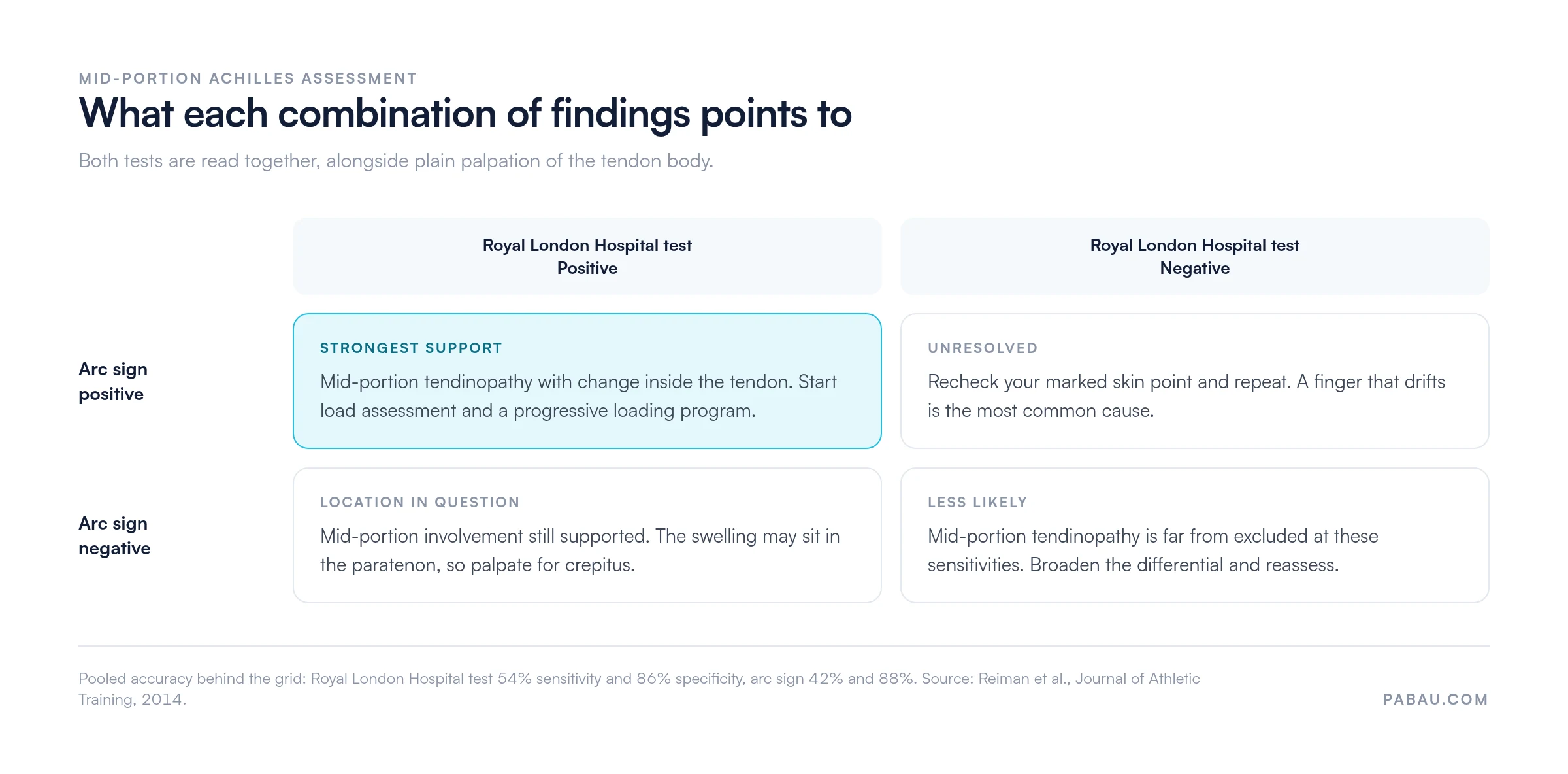
Record which test was positive rather than a single “RLH positive” note. The two findings answer different questions, and a colleague reading your note later cannot reconstruct the difference from one line. Practice management software like Pabau gives each result its own field, alongside the patient’s own words and the working differential. It all sits in one structured record the whole team can open.
Clear documentation at assessment also supports onward referral when imaging is warranted. A note that records each test separately helps findings travel accurately between practitioners.

A negative battery lowers the probability of mid-portion tendinopathy without ruling it out. When both tests come back negative, these differentials deserve attention.
Only two primary studies have measured these tests against imaging or histology, and both are small. Maffulli et al. examined 10 male athletes and 14 controls in 2003. Hutchison et al. examined 21 patients in 2013. Reiman et al. pooled the two in 2014, giving a combined sample of 45.
Specificity is the useful half of the picture. A positive Royal London Hospital test raises the odds of mid-portion tendinopathy by roughly four times. That is enough to act on in a patient who already fits the clinical picture. Sensitivity near 0.5 means a negative result changes very little, so it should never end the assessment.
Combining the three tests did not beat the Royal London Hospital test on its own in the Maffulli data. The battery reached 83% specificity against 91% for the single test. Its value lies in telling you where the lesion sits and which tissue is involved.
Reliability differed between the two studies too. Maffulli et al. reported interrater kappa of 0.63 to 0.76 for the Royal London Hospital test. Hutchison et al. reported 0.37 for the same test. Treat a result from an unfamiliar examiner with appropriate caution.
Note the ankle angle at which tenderness drops away, not just the fact that it did. Tenderness that only settles in the last few degrees of dorsiflexion behaves differently at reassessment from tenderness that vanishes at mid-range. Recording the angle gives you a comparison point six weeks later that a simple positive or negative cannot provide.
Each test brings something the other does not, and each has firm limits. Knowing where those limits fall prevents over-reading a result.
A knee version does exist, and it is the source of most of the confusion. Maffulli and colleagues published a Royal London Hospital test for patellar tendinopathy in 2017, applying the same logic to a different tendon.
There, the examiner finds the tender point with the knee extended, then palpates it again with the knee flexed to 90 degrees. Pain that is markedly reduced or absent in flexion counts as positive. Against ultrasound the test reached 88% sensitivity and 98% specificity in that cohort.
The two tests share a name and a principle, and little else. Citing the patellar figures in an Achilles assessment, or the reverse, misstates the evidence. Check which tendon a quoted sensitivity belongs to before it reaches a patient record.
Neither test should drive management on its own. They sit inside a wider assessment that measures severity, capacity and irritability. Here is what they pair with.
For athletes returning to sport, a structured return-to-running protocol should start only once tenderness is minimal and single-leg calf raises are comfortable. Practices using a physical therapy EMR can tie stage-progression reminders to the reassessment schedule, which reduces the risk of an early return.
In most physical therapy practices, a tendon assessment is typed as free text. Six weeks later nobody can tell whether the arc sign was tested and negative, or simply never performed. Reassessment then starts from scratch, and progress is judged on memory.
Pabau replaces that with a structured form. Each test gets its own field, so a negative result is recorded as a negative rather than an absence. The marked palpation point, the dorsiflexion angle and the patient’s own description sit in the same note, ready to compare at the next visit.
From there the record does some work for you. Reassessment recalls go out automatically on the interval you set, and VISA-A scores are captured through the patient portal before the appointment. Every clinician who opens the file sees the same assessment history. The result is a tendinopathy pathway your whole team runs the same way.

Pabau gives physical therapy and sports medicine practices one platform for digital intake forms, structured clinical notes, and automated patient recalls. See how it fits your assessment workflow.

The Royal London Hospital test and the arc sign are two tests, not two names for one. The first asks whether tenderness fades when the tendon is put under tension. The second asks whether the swelling travels with the tendon. Run them together with palpation and you get a location and a tissue in under three minutes.
Keep the accuracy in proportion. A positive result is worth acting on, a negative result settles very little, and the evidence behind both rests on 45 participants. Pair the battery with load testing, a patient-reported score and imaging where the picture stays unclear.
Pabau helps physical therapy and sports medicine practices standardize this pathway, record each test result separately, and schedule reassessments automatically. Book a demo to see how that keeps an Achilles pathway consistent across your whole team.

Need to rule out a rupture first? Thompson test walks through the calf-squeeze maneuver and what a normal plantarflexion response looks like.
Managing a confirmed rupture? Achilles tendon rupture treatment guidelines handout gives you a patient-facing summary of the conservative and surgical pathways.
Need to clear a bony injury? Ottawa ankle rules calculator sets out the four palpation points that decide whether an X-ray is warranted.
Suspect the pain is bony rather than tendinous? Foot stress fracture test covers the hop, tuning fork and percussion tests and their reported accuracy.
Building a loading program for another tendon? Exercises for gluteal tendinopathy handout gives patients a staged routine you can adapt to your own reassessment intervals.
The Royal London Hospital test helps confirm mid-portion Achilles tendinopathy at the bedside. The examiner finds the tender spot with the ankle resting in plantarflexion, then palpates the same spot again in maximal dorsiflexion. A positive result is tenderness that disappears, or drops sharply, in that dorsiflexed position.
No. They are two distinct tests that Maffulli et al. described together in 2003. The arc sign is positive when the tender, swollen area of tendon moves with the tendon as the ankle is dorsiflexed and plantarflexed. The Royal London Hospital test is positive when tenderness at a fixed spot falls away in dorsiflexion. Clinicians normally run both, alongside plain palpation, as one battery.
Reiman et al. pooled two studies in 2014 and reported 54% sensitivity (95% CI 34-73%) with 86% specificity (95% CI 72-95%). In the original Maffulli et al. 2003 cohort, the test alone gave 54% sensitivity and 91% specificity. Palpation, the arc sign and the test combined reached 59% sensitivity and 83% specificity in that study.
A positive arc sign points to pathology inside the tendon body rather than around it. Swelling that travels with the tendon during ankle movement sits within the tendon itself. Swelling that stays fixed while the tendon slides under it suggests paratendinopathy instead. Pooled accuracy for the arc sign is 42% sensitivity and 88% specificity.
Yes, but it is a separate test for a separate tendon. Maffulli et al. described a patellar version in 2017, palpating the tender spot with the knee extended and again at 90 degrees of flexion. Against ultrasound it reached 88% sensitivity and 98% specificity. The Achilles figures do not transfer to the knee, and the knee figures do not transfer to the Achilles.
No. Both studies in the Reiman review recruited patients with mid-portion disease, 2-6 cm above the calcaneal insertion. Insertional pathology is anchored at the calcaneal attachment, so tenderness there tends to persist or worsen in dorsiflexion. No published accuracy figures support using either test for insertional tendinopathy.