CRM for chiropractors: how to manage patients, bookings and marketing
A CRM for chiropractors is a patient relationship management system built around...

Last Updated: Oct 13, 2026

The Matles test is a bedside check for complete Achilles tendon rupture, performed with the patient prone and knees actively flexed to 90 degrees.
A positive result means the affected foot falls to neutral or into dorsiflexion instead of holding slight plantarflexion, which indicates loss of tendon continuity.
Reported sensitivity is 88% and specificity 85% for complete rupture, based on Maffulli 1998, but the test misses partial tears.
Used alongside the Thompson (Simmonds) test, the Matles test strengthens clinical confidence before imaging is ordered.
Missing an Achilles tendon rupture at first presentation is one of the more consequential errors in musculoskeletal medicine. Swelling and pain can mimic an ankle sprain. Many patients also keep some active plantarflexion through intact accessory muscles, so the injury looks milder than it is.
The Matles test gives you a passive bedside check that does not depend on patient effort or pain tolerance. This guide covers the anatomy behind it, the step-by-step technique, and how to read the result. It also sets the published accuracy figures against the Thompson test, so you can use the two signs together with confidence.
The Matles test is a passive clinical examination used to assess the integrity of the Achilles tendon. It exploits the resting tension of an intact tendon. When the knee is flexed to 90 degrees in a prone patient, the gastrocnemius-soleus complex normally holds the foot in slight plantarflexion. If the tendon is ruptured, that resting tension is lost and the foot drifts toward neutral or into dorsiflexion.
The test is particularly useful in the acute setting, where swelling and pain limit active movement testing. Because it relies on passive observation rather than active patient effort, the result is not confounded by guarding or incomplete effort. It was described as a complement to the Thompson (Simmonds) test rather than a replacement, and the evidence still supports using them together.
The Achilles tendon is the largest and strongest tendon in the body. It is formed by the conjoined tendons of the gastrocnemius and soleus muscles, collectively the triceps surae, and inserts into the posterior calcaneus. It transmits force from the calf complex to the heel, producing plantarflexion at the ankle and propulsion during gait.
The tendon has a zone of relative avascularity roughly 2 to 6 centimeters proximal to the calcaneal insertion, and that is where most ruptures occur. While the tendon is intact, passive elastic tension holds the foot in plantarflexion at rest. With the knee at 90 degrees, that is roughly 20 to 30 degrees. Rupture eliminates that tension.
The positional change the Matles test detects follows directly from that mechanism. Understanding it is what lets you interpret a result in a patient with a partial tear, where residual tendon fibers still preserve some resting tension.
Technique is straightforward, but bilateral comparison is essential. Always examine the unaffected limb first to establish the patient’s normal resting foot position.
The whole test takes under 60 seconds, and no equipment is required beyond an examination table. The prone position may be uncomfortable for a patient with acute calf pain or hematoma. In that case, note the limitation and use the Thompson test as the primary screen.
Interpretation is bilateral and comparative, not absolute. The affected side is always read against the contralateral limb. Clinicians sometimes call the dropped resting position the “angle of dangle.”
One clinical point is easy to miss. Some patients with complete rupture retain active plantarflexion through the peroneus longus and tibialis posterior. The Matles test bypasses active effort and reads passive resting tension instead. That is why it can pick up a rupture even when the patient plantarflexes on command.
The primary diagnostic accuracy data comes from Maffulli et al. (1998, American Journal of Sports Medicine). It remains the most widely cited study on bedside clinical tests for Achilles tendon rupture. The research evaluated multiple clinical signs in patients with surgically confirmed complete ruptures.
These figures apply to complete Achilles tendon rupture, and they cannot be extrapolated to partial tears. Maffulli’s cohort was made up of surgically confirmed cases, so the numbers reflect favorable examination conditions. They are not emergency department presentations with early hematoma and limited access to the limb. Expect real-world sensitivity to sit slightly lower.
The Thompson test, also called the Simmonds test or calf squeeze test, is the most commonly used bedside screen for Achilles tendon rupture. Knowing how it differs from the Matles test helps you choose the right test for the situation in front of you. It also explains why the pair outperforms either sign alone.
Neither test alone rules out rupture. What matters clinically is the combination, and the grid below sets out the four ways the two results can land.
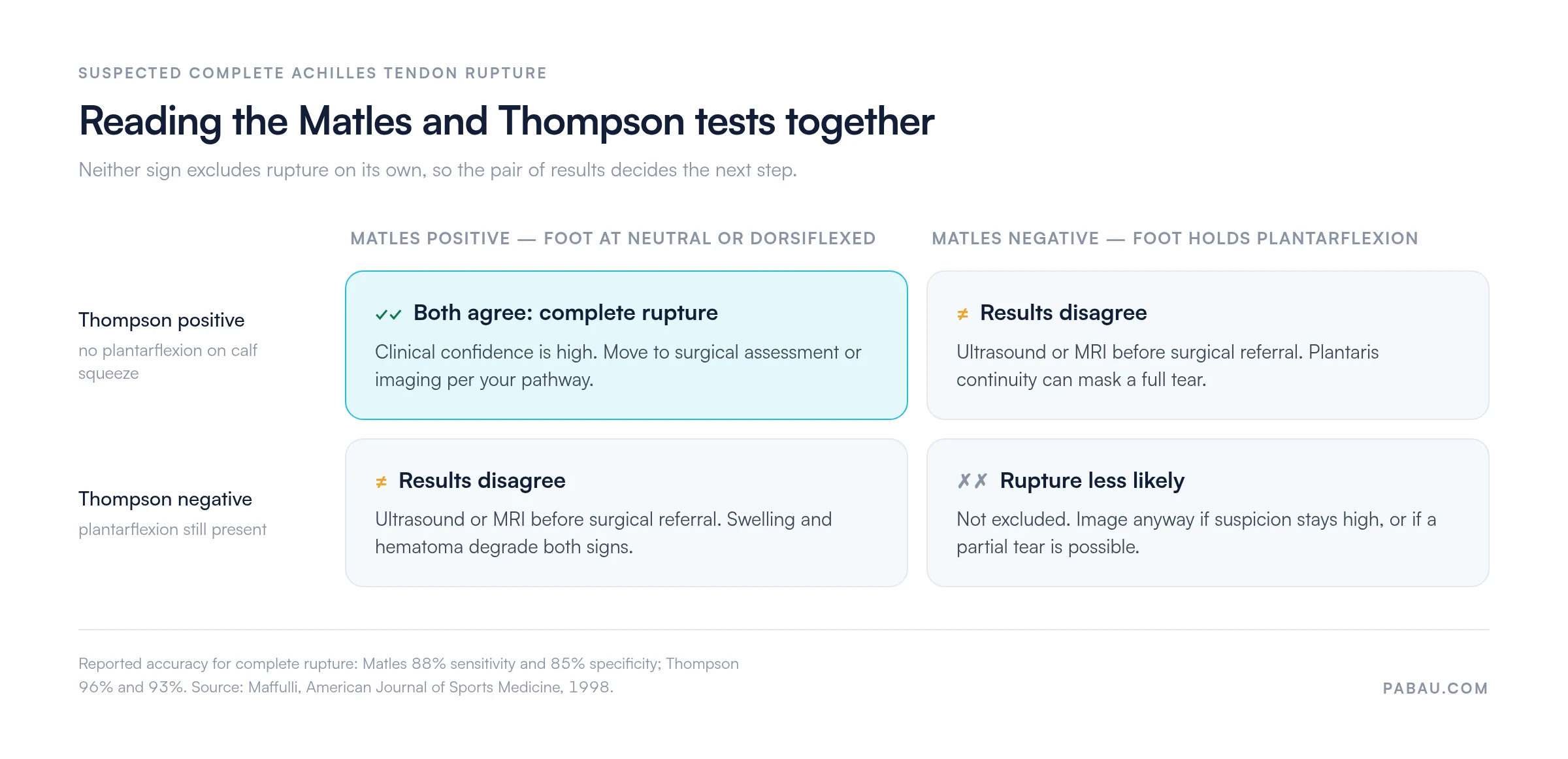
The Matles test adds the most value when the Thompson test is equivocal, or when calf palpation is too painful to be reliable. In a delayed presentation, more than 72 hours after injury, organizing hematoma and swelling can blunt the Thompson test. The resting-position approach of the Matles test becomes relatively more informative at that point.
The Matles test is not a universal screen for ankle pain. It has a specific indication: suspected complete Achilles tendon rupture in a patient with sudden posterior heel or lower calf pain. Patients often describe a “snap” or a “kick” sensation, with or without an audible pop. Deep buttock or posterior thigh pain points to the proximal hamstring instead, which the Puranen-Orava test screens.
Where Achilles rupture sits in the differential for an ankle or foot injury, start with the Ottawa ankle rules. They tell you whether plain radiographs are indicated. Once fracture is excluded, clinical tests like the Matles test carry far more weight.
Teams also need a plan for what happens after the diagnosis. A structured return-to-running protocol governs loading once the rupture is confirmed and treated. Purpose-built software for physical therapists keeps bilateral findings in structured fields rather than free text, so the angles stay retrievable at follow-up.
Knowing where a test fails matters as much as knowing where it succeeds. The Matles test has several limitations that cap how much weight a negative result can carry.
When the Matles test is negative but clinical suspicion remains, ultrasound is the preferred next step. It is highly sensitive for complete rupture, works dynamically during the exam, and involves no ionizing radiation. For the pathway that follows a confirmed rupture, our Achilles rupture treatment guidelines handout is a printable reference to keep with the patient’s notes.
A free-text note usually records the Matles test as a single word: positive or negative. The degree of asymmetry, the side, and the comparison with the previous visit all disappear into prose. Six weeks later, nobody can say whether the resting foot position has changed.
Practice management software like Pabau lets you build the examination as a structured template instead. Bilateral Matles and Thompson results get their own fields, so every clinician on the team records the same measurements in the same place. Photographs of the resting foot position attach to the same encounter.
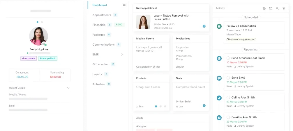
The payoff shows up at the follow-up visit and in the referral letter. You can pull the initial angles, compare them with today’s, and hand the orthopedic team a documented sequence instead of a recollection. Pabau GO, our iOS app, opens the same client record on a phone at the bedside.

Pabau’s structured examination templates capture Matles and Thompson results side by side in the client record, with photographs attached to the same visit. Findings stay retrievable for follow-ups, referral letters, and outcome tracking.

The Matles test earns its place as the second opinion in an examination that should never rest on one sign. Run the Thompson test first, then the Matles test, and read the two together. Two results that agree move you toward referral, and two that disagree move you toward ultrasound.
The trade-off worth remembering is that a negative result buys you very little. Plantaris continuity and partial tears both produce false negatives, so clinical suspicion outranks the sign. Record the degrees on each side rather than a verdict, and the next clinician can check your reasoning. Book a demo to see how Pabau keeps bilateral examination findings in one client record.

Need to rule out a fracture first? Ottawa ankle rules calculator shows when plain radiographs are indicated before you move on to soft-tissue assessment.
Planning the return to sport after repair? Return-to-running protocol for physical therapy covers progressive loading criteria and clearance milestones after a lower limb tendon injury.
Need the treatment pathway on paper? Achilles tendon rupture treatment guidelines handout is a printable reference you can keep with the patient’s notes.
Working through posterior foot and heel pain? Foot stress fracture test walks through the bedside assessment that points to bone rather than tendon.
The Matles test is a bedside clinical examination used to assess whether the Achilles tendon is intact or ruptured. It is indicated for suspected complete rupture. The examiner watches the resting foot position while the patient holds both knees actively flexed to 90 degrees, lying prone.
A positive Matles test means the affected foot drifts to neutral or falls into dorsiflexion when the knee is flexed to 90 degrees. The unaffected side holds its normal slight plantarflexion. That loss of resting tone points to a complete rupture of the Achilles tendon, where passive calf tension is gone.
Position the patient prone with both feet hanging free. Ask them to actively flex both knees to 90 degrees, then observe both feet at once without touching them. Compare the resting foot angle bilaterally. A normal foot rests in slight plantarflexion, so a foot at neutral or in dorsiflexion is positive.
Maffulli et al. reported a sensitivity of 88% and a specificity of 85% for complete Achilles tendon rupture. That study appeared in the American Journal of Sports Medicine in 1998. The figures cover complete rupture only, and the test is not reliable for partial tears.
In the Thompson (Simmonds) test the examiner squeezes the calf and watches for plantarflexion. The Matles test uses the patient’s own knee flexion and passive observation of resting foot position. The Thompson test is slightly more sensitive, at 96% against 88%. The two complement each other, so agreeing results give strong confidence and discordant results call for imaging.
No. The Matles test is not reliable for partial Achilles tears. Residual tendon fibers in a partial rupture preserve some resting plantarflexion tension, which produces a false negative. Where a partial tear is suspected, ultrasound is the appropriate next investigation rather than a bedside test.